5JQR

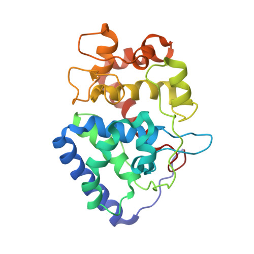
The Structure of Ascorbate Peroxidase Compound II formed by reaction with m-CPBA


Experimental Data Snapshot

  • Method: X-RAY DIFFRACTION
  • Resolution: 1.81 Å
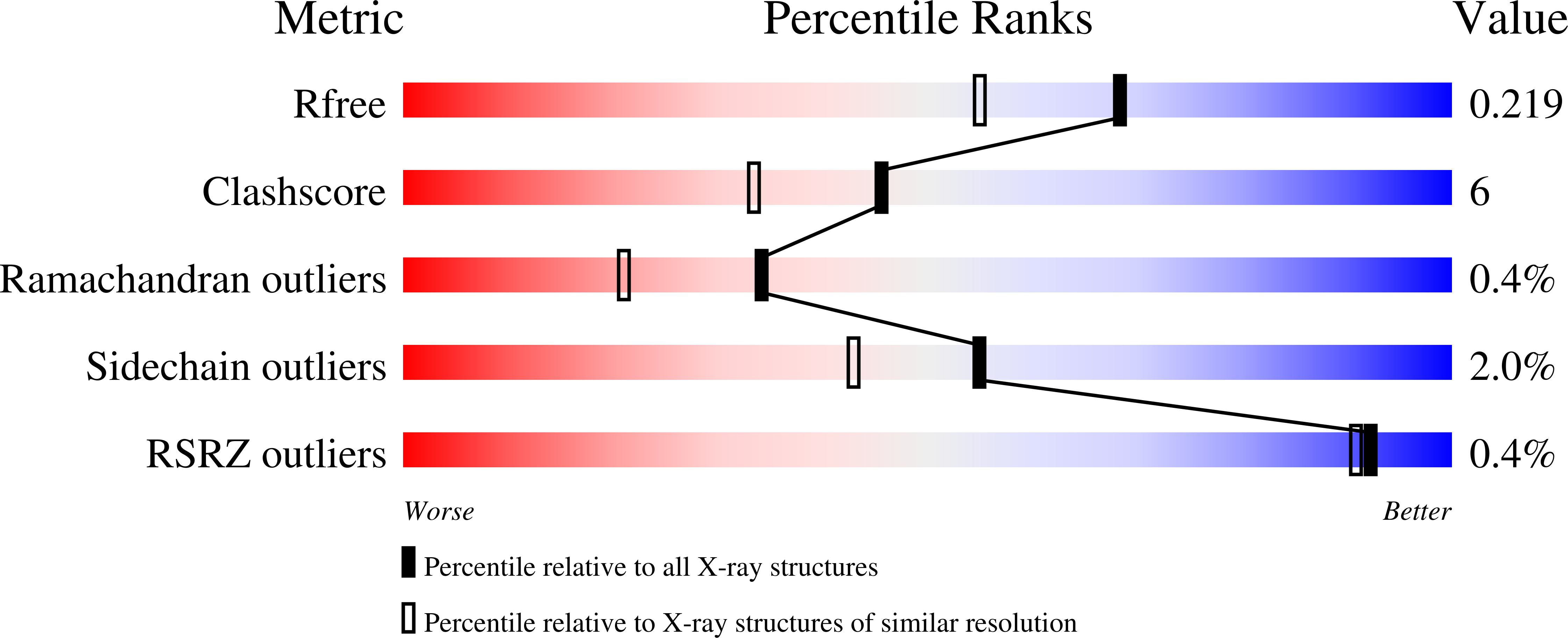
  • R-Value Free: 0.214 
  • R-Value Work: 0.154 
  • R-Value Observed: 0.157 

Starting Model: experimental
View more details

wwPDB Validation   3D Report Full Report


Ligand Structure Quality Assessment 


This is version 2.1 of the entry. See complete history


Literature

Direct visualization of a Fe(IV)-OH intermediate in a heme enzyme.

Kwon, H.Basran, J.Casadei, C.M.Fielding, A.J.Schrader, T.E.Ostermann, A.Devos, J.M.Aller, P.Blakeley, M.P.Moody, P.C.Raven, E.L.

(2016) Nat Commun 7: 13445-13445

  • DOI: https://doi.org/10.1038/ncomms13445
  • Primary Citation of Related Structures:  
    5JPR, 5JQR

  • PubMed Abstract: 

    Catalytic heme enzymes carry out a wide range of oxidations in biology. They have in common a mechanism that requires formation of highly oxidized ferryl intermediates. It is these ferryl intermediates that provide the catalytic engine to drive the biological activity. Unravelling the nature of the ferryl species is of fundamental and widespread importance. The essential question is whether the ferryl is best described as a Fe(IV)=O or a Fe(IV)-OH species, but previous spectroscopic and X-ray crystallographic studies have not been able to unambiguously differentiate between the two species. Here we use a different approach. We report a neutron crystal structure of the ferryl intermediate in Compound II of a heme peroxidase; the structure allows the protonation states of the ferryl heme to be directly observed. This, together with pre-steady state kinetic analyses, electron paramagnetic resonance spectroscopy and single crystal X-ray fluorescence, identifies a Fe(IV)-OH species as the reactive intermediate. The structure establishes a precedent for the formation of Fe(IV)-OH in a peroxidase.


  • Organizational Affiliation

    Department of Molecular and Cell Biology and Leicester Institute of Structural and Chemical Biology, University of Leicester, Lancaster Road, Leicester LE1 9HN, UK.


Macromolecules
Find similar proteins by:  (by identity cutoff)  |  3D Structure
Entity ID: 1
MoleculeChains Sequence LengthOrganismDetailsImage
Ascorbate peroxidase249Glycine maxMutation(s): 0 
Gene Names: apx1GLYMA_U021900
EC: 1.11.1.11
UniProt
Find proteins for Q43758 (Glycine max)
Explore Q43758 
Go to UniProtKB:  Q43758
Entity Groups  
Sequence Clusters30% Identity50% Identity70% Identity90% Identity95% Identity100% Identity
UniProt GroupQ43758
Sequence Annotations
Expand
  • Reference Sequence
Experimental Data & Validation

Experimental Data

  • Method: X-RAY DIFFRACTION
  • Resolution: 1.81 Å
  • R-Value Free: 0.214 
  • R-Value Work: 0.154 
  • R-Value Observed: 0.157 
  • Space Group: P 42 21 2
Unit Cell:
Length ( Å )Angle ( ˚ )
a = 82.095α = 90
b = 82.095β = 90
c = 75.162γ = 90
Software Package:
Software NamePurpose
REFMACrefinement
iMOSFLMdata reduction
Aimlessdata scaling
PHASERphasing

Structure Validation

View Full Validation Report


Ligand Structure Quality Assessment 


Entry History & Funding Information

Deposition Data


Funding OrganizationLocationGrant Number
Biotechnology and Biological Sciences Research CouncilUnited KingdomBB/K015656/1
Wellcome TrustUnited KingdomWT094104MA

Revision History  (Full details and data files)

  • Version 1.0: 2016-12-21
    Type: Initial release
  • Version 2.0: 2017-08-30
    Changes: Atomic model, Author supporting evidence
  • Version 2.1: 2024-01-10
    Changes: Data collection, Database references, Derived calculations, Refinement description