5JQV

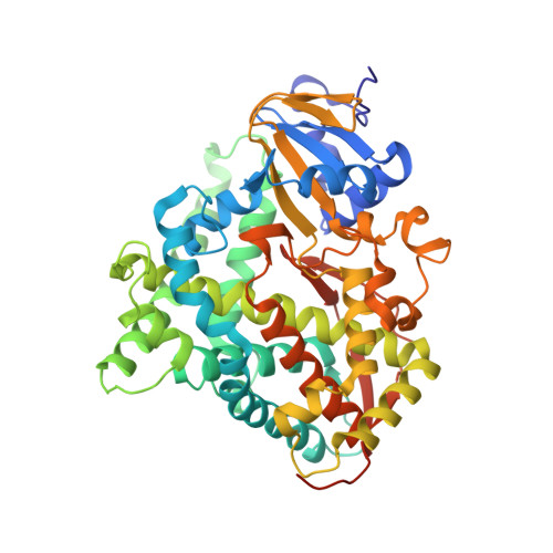
Crystal structure of Cytochrome P450 BM3 heme domain T269V/L272W/L322I/A406S (WIVS) variant with iron(III) deuteroporphyrin IX bound


Experimental Data Snapshot

  • Method: X-RAY DIFFRACTION
  • Resolution: 2.34 Å
  • R-Value Free: 0.284 
  • R-Value Work: 0.215 
  • R-Value Observed: 0.215 

Starting Model: experimental
View more details

wwPDB Validation   3D Report Full Report


Ligand Structure Quality Assessment 


This is version 1.4 of the entry. See complete history


Literature

An Evolved Orthogonal Enzyme/Cofactor Pair.

Reynolds, E.W.McHenry, M.W.Cannac, F.Gober, J.G.Snow, C.D.Brustad, E.M.

(2016) J Am Chem Soc 138: 12451-12458

  • DOI: https://doi.org/10.1021/jacs.6b05847
  • Primary Citation of Related Structures:  
    5JQU, 5JQV

  • PubMed Abstract: 

    We introduce a strategy that expands the functionality of hemoproteins through orthogonal enzyme/heme pairs. By exploiting the ability of a natural heme transport protein, ChuA, to promiscuously import heme derivatives, we have evolved a cytochrome P450 (P450BM3) that selectively incorporates a nonproteinogenic cofactor, iron deuteroporphyrin IX (Fe-DPIX), even in the presence of endogenous heme. Crystal structures show that selectivity gains are due to mutations that introduce steric clash with the heme vinyl groups while providing a complementary binding surface for the smaller Fe-DPIX cofactor. Furthermore, the evolved orthogonal enzyme/cofactor pair is active in non-natural carbenoid-mediated olefin cyclopropanation. This methodology for the generation of orthogonal enzyme/cofactor pairs promises to expand cofactor diversity in artificial metalloenzymes.


  • Organizational Affiliation

    Department of Chemistry, University of North Carolina-Chapel Hill , 125 South Road, CB 3290, Chapel Hill, North Carolina 27599, United States.


Macromolecules
Find similar proteins by:  (by identity cutoff)  |  3D Structure
Entity ID: 1
MoleculeChains Sequence LengthOrganismDetailsImage
Bifunctional cytochrome P450/NADPH--P450 reductase
A, B, C, D, E
A, B, C, D, E, F, G, H
471Priestia megaterium NBRC 15308 = ATCC 14581Mutation(s): 4 
Gene Names: cyp102A1cyp102BG04_163
EC: 1.14.14.1 (PDB Primary Data), 1.6.2.4 (PDB Primary Data)
UniProt
Find proteins for P14779 (Priestia megaterium (strain ATCC 14581 / DSM 32 / CCUG 1817 / JCM 2506 / NBRC 15308 / NCIMB 9376 / NCTC 10342 / NRRL B-14308 / VKM B-512 / Ford 19))
Explore P14779 
Go to UniProtKB:  P14779
Entity Groups  
Sequence Clusters30% Identity50% Identity70% Identity90% Identity95% Identity100% Identity
UniProt GroupP14779
Sequence Annotations
Expand
  • Reference Sequence
Experimental Data & Validation

Experimental Data

  • Method: X-RAY DIFFRACTION
  • Resolution: 2.34 Å
  • R-Value Free: 0.284 
  • R-Value Work: 0.215 
  • R-Value Observed: 0.215 
  • Space Group: P 21 21 21
Unit Cell:
Length ( Å )Angle ( ˚ )
a = 106.708α = 90
b = 167.405β = 90
c = 228.471γ = 90
Software Package:
Software NamePurpose
PHENIXrefinement
PDB_EXTRACTdata extraction
PHASERphasing

Structure Validation

View Full Validation Report


Ligand Structure Quality Assessment 


Entry History & Funding Information

Deposition Data


Funding OrganizationLocationGrant Number
Defense Advanced Research Projects AgencyUnited StatesD13AP00024
National Science Foundation (NSF, United States)United StatesDGE1144081

Revision History  (Full details and data files)

  • Version 1.0: 2016-09-28
    Type: Initial release
  • Version 1.1: 2016-10-05
    Changes: Database references
  • Version 1.2: 2017-09-20
    Changes: Author supporting evidence, Derived calculations
  • Version 1.3: 2019-11-27
    Changes: Author supporting evidence
  • Version 1.4: 2023-09-27
    Changes: Data collection, Database references, Derived calculations, Refinement description