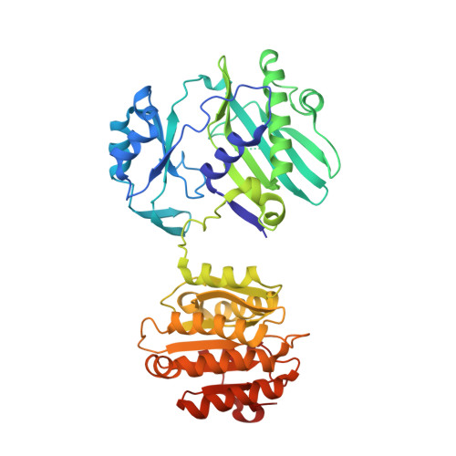
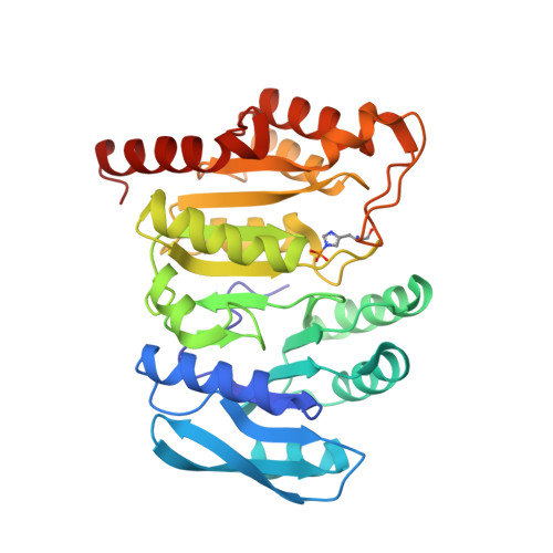
Binding of hydroxycitrate to human ATP-citrate lyase.
Hu, J., Komakula, A., Fraser, M.E.(2017) Acta Crystallogr D Struct Biol 73: 660-671
- PubMed: 28777081
- DOI: https://doi.org/10.1107/S2059798317009871
- Primary Citation of Related Structures:
5TDE, 5TDF, 5TDM, 5TDZ, 5TE1, 5TEQ, 5TES, 5TET - PubMed Abstract:
Hydroxycitrate from the fruit of Garcinia cambogia [i.e. (2S,3S)-2-hydroxycitrate] is the best-known inhibitor of ATP-citrate lyase. Well diffracting crystals showing how the inhibitor binds to human ATP-citrate lyase were grown by modifying the protein. The protein was modified by introducing cleavage sites for Tobacco etch virus protease on either side of a disordered linker. The protein crystallized consisted of residues 2-425-ENLYFQ and S-488-810 of human ATP-citrate lyase. (2S,3S)-2-Hydroxycitrate binds in the same orientation as citrate, but the citrate-binding domain (residues 248-421) adopts a different orientation with respect to the rest of the protein (residues 4-247, 490-746 and 748-809) from that previously seen. For the first time, electron density was evident for the loop that contains His760, which is phosphorylated as part of the catalytic mechanism. The pro-S carboxylate of (2S,3S)-2-hydroxycitrate is available to accept a phosphoryl group from His760. However, when co-crystals were grown with ATP and magnesium ions as well as either the inhibitor or citrate, Mg 2+ -ADP was bound and His760 was phosphorylated. The phosphoryl group was not transferred to the organic acid. This led to the interpretation that the active site is trapped in an open conformation. The strategy of designing cleavage sites to remove disordered residues could be useful in determining the crystal structures of other proteins.
Organizational Affiliation:
Department of Biological Sciences, University of Calgary, 2500 University Drive NW, Calgary, Alberta T2N 1N4, Canada.


