5Y0B

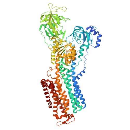
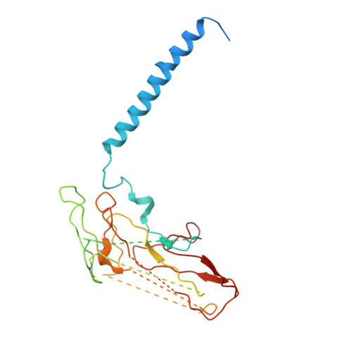
PIG GASTRIC H+,K+ - ATPASE IN COMPLEX with BYK99


Experimental Data Snapshot

  • Method: ELECTRON CRYSTALLOGRAPHY
  • Resolution: 6.50 Å

wwPDB Validation   3D Report Full Report


This is version 1.1 of the entry. See complete history


Literature

The cryo-EM structure of gastric H(+),K(+)-ATPase with bound BYK99, a high-affinity member of K(+)-competitive, imidazo[1,2-a]pyridine inhibitors

Abe, K.Shimokawa, J.Naito, M.Munson, K.Vagin, O.Sachs, G.Suzuki, H.Tani, K.Fujiyoshi, Y.

(2017) Sci Rep 7: 6632-6632

  • DOI: https://doi.org/10.1038/s41598-017-06698-8
  • Primary Citation of Related Structures:  
    5Y0B

  • PubMed Abstract: 

    The gastric proton pump H + ,K + -ATPase acidifies the gastric lumen, and thus its inhibitors, including the imidazo[1,2-a]pyridine class of K + -competitive acid blockers (P-CABs), have potential application as acid-suppressing drugs. We determined the electron crystallographic structure of H + ,K + -ATPase at 6.5 Å resolution in the E2P state with bound BYK99, a potent P-CAB with a restricted ring structure. The BYK99 bound structure has an almost identical profile to that of a previously determined structure with bound SCH28080, the original P-CAB prototype, but is significantly different from the previously reported P-CAB-free form, illustrating a common conformational change is required for P-CAB binding. The shared conformational changes include a distinct movement of transmembrane helix 2 (M2), from its position in the previously reported P-CAB-free form, to a location proximal to the P-CAB binding site in the present BYK99-bound structure. Site-specific mutagenesis within M2 revealed that D137 and N138, which face the P-CAB binding site in our model, significantly affect the inhibition constant (K i ) of P-CABs. We also found that A335 is likely to be near the bridging nitrogen at the restricted ring structure of the BYK99 inhibitor. These provide clues to elucidate the binding site parameters and mechanism of P-CAB inhibition of gastric acid secretion.


  • Organizational Affiliation

    Graduate School of Pharmaceutical Sciences, Nagoya University, Nagoya, 464-8601, Japan. kabe@cespi.nagoya-u.ac.jp.


Macromolecules
Find similar proteins by:  (by identity cutoff)  |  3D Structure
Entity ID: 1
MoleculeChains Sequence LengthOrganismDetailsImage
Potassium-transporting ATPase alpha chain 11,034Sus scrofaMutation(s): 0 
EC: 3.6.3.10 (PDB Primary Data), 7.2.2.19 (UniProt)
Membrane Entity: Yes 
UniProt
Find proteins for P19156 (Sus scrofa)
Explore P19156 
Go to UniProtKB:  P19156
Entity Groups  
Sequence Clusters30% Identity50% Identity70% Identity90% Identity95% Identity100% Identity
UniProt GroupP19156
Sequence Annotations
Expand
  • Reference Sequence
Find similar proteins by:  (by identity cutoff)  |  3D Structure
Entity ID: 2
MoleculeChains Sequence LengthOrganismDetailsImage
Potassium-transporting ATPase subunit beta290Sus scrofaMutation(s): 0 
Membrane Entity: Yes 
UniProt
Find proteins for P18434 (Sus scrofa)
Explore P18434 
Go to UniProtKB:  P18434
Entity Groups  
Sequence Clusters30% Identity50% Identity70% Identity90% Identity95% Identity100% Identity
UniProt GroupP18434
Sequence Annotations
Expand
  • Reference Sequence
Experimental Data & Validation

Experimental Data

  • Method: ELECTRON CRYSTALLOGRAPHY
  • Resolution: 6.50 Å
  • Space Group: P 21 21 2
Unit Cell:
Length ( Å )Angle ( ˚ )
a = 142.6α = 90
b = 112β = 90
c = 320γ = 90
EM Software:
TaskSoftware PackageVersion
RECONSTRUCTIONMRC
MODEL REFINEMENTCoot

Structure Validation

View Full Validation Report


Entry History & Funding Information

Deposition Data


Funding OrganizationLocationGrant Number
Japan--

Revision History  (Full details and data files)

  • Version 1.0: 2017-08-09
    Type: Initial release
  • Version 1.1: 2024-11-06
    Changes: Data collection, Database references, Refinement description, Structure summary