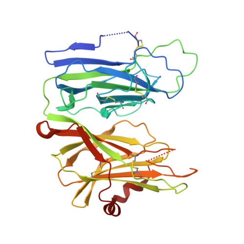
6AMP

Crystal structure of H172A PHM (CuH absent, CuM present)


Experimental Data Snapshot

  • Method: X-RAY DIFFRACTION
  • Resolution: 2.48 Å
  • R-Value Free: 0.290 
  • R-Value Work: 0.213 
  • R-Value Observed: 0.217 

Starting Model: experimental
View more details

wwPDB Validation   3D Report Full Report


This is version 1.3 of the entry. See complete history


Literature

Effects of copper occupancy on the conformational landscape of peptidylglycine alpha-hydroxylating monooxygenase.

Maheshwari, S.Shimokawa, C.Rudzka, K.Kline, C.D.Eipper, B.A.Mains, R.E.Gabelli, S.B.Blackburn, N.Amzel, L.M.

(2018) Commun Biol 1: 74-74

  • DOI: https://doi.org/10.1038/s42003-018-0082-y
  • Primary Citation of Related Structures:  
    5WJA, 5WKW, 5WM0, 6ALA, 6ALV, 6AMP, 6AN3, 6AO6, 6AY0

  • PubMed Abstract: 

    The structures of metalloproteins that use redox-active metals for catalysis are usually exquisitely folded in a way that they are prearranged to accept their metal cofactors. Peptidylglycine α-hydroxylating monooxygenase (PHM) is a dicopper enzyme that catalyzes hydroxylation of the α-carbon of glycine-extended peptides for the formation of des-glycine amidated peptides. Here, we present the structures of apo-PHM and of mutants of one of the copper sites (H107A, H108A, and H172A) determined in the presence and absence of citrate. Together, these structures show that the absence of one copper changes the conformational landscape of PHM. In one of these structures, a large interdomain rearrangement brings residues from both copper sites to coordinate a single copper (closed conformation) indicating that full copper occupancy is necessary for locking the catalytically competent conformation (open). These data suggest that in addition to their required participation in catalysis, the redox-active metals play an important structural role.


  • Organizational Affiliation

    Department of Biophysics and Biophysical Chemistry, Johns Hopkins University School of Medicine, Baltimore, MD, 21205, USA.


Macromolecules
Find similar proteins by:  (by identity cutoff)  |  3D Structure
Entity ID: 1
MoleculeChains Sequence LengthOrganismDetailsImage
Peptidyl-glycine alpha-amidating monooxygenase312Rattus norvegicusMutation(s): 1 
Gene Names: Pam
EC: 1.14.17.3 (PDB Primary Data), 4.3.2.5 (PDB Primary Data)
UniProt
Find proteins for P14925 (Rattus norvegicus)
Explore P14925 
Go to UniProtKB:  P14925
Entity Groups  
Sequence Clusters30% Identity50% Identity70% Identity90% Identity95% Identity100% Identity
UniProt GroupP14925
Sequence Annotations
Expand
  • Reference Sequence
Small Molecules
Ligands 1 Unique
IDChains Name / Formula / InChI Key2D Diagram3D Interactions
CU
Query on CU

Download Ideal Coordinates CCD File 
B [auth A]COPPER (II) ION
Cu
JPVYNHNXODAKFH-UHFFFAOYSA-N
Experimental Data & Validation

Experimental Data

  • Method: X-RAY DIFFRACTION
  • Resolution: 2.48 Å
  • R-Value Free: 0.290 
  • R-Value Work: 0.213 
  • R-Value Observed: 0.217 
  • Space Group: P 21 21 21
Unit Cell:
Length ( Å )Angle ( ˚ )
a = 59.422α = 90
b = 66.362β = 90
c = 69.79γ = 90
Software Package:
Software NamePurpose
REFMACrefinement
DENZOdata collection
SCALEPACKdata scaling
PDB_EXTRACTdata extraction
Cootmodel building
SCALEPACKdata reduction
REFMACphasing

Structure Validation

View Full Validation Report


Entry History & Funding Information

Deposition Data


Funding OrganizationLocationGrant Number
National Science Foundation (NSF, United States)United StatesMCB-1517522

Revision History  (Full details and data files)

  • Version 1.0: 2018-07-18
    Type: Initial release
  • Version 1.1: 2018-10-17
    Changes: Data collection, Database references
  • Version 1.2: 2019-11-27
    Changes: Author supporting evidence
  • Version 1.3: 2024-04-03
    Changes: Data collection, Database references, Refinement description