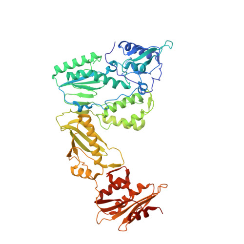
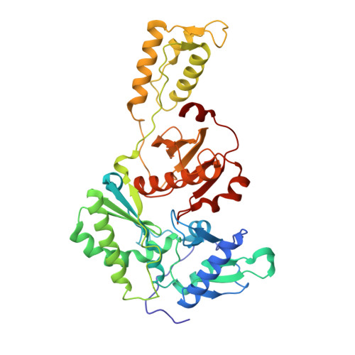
6AOC

Crystal Structure of an N-Hydroxythienopyrimidine-2,4-dione RNase H Active Site Inhibitor with Multiple Binding Modes to HIV Reverse Transcriptase


Experimental Data Snapshot

  • Method: X-RAY DIFFRACTION
  • Resolution: 1.80 Å
  • R-Value Free: 0.226 
  • R-Value Work: 0.186 
  • R-Value Observed: 0.188 

Starting Model: experimental
View more details

wwPDB Validation   3D Report Full Report


Ligand Structure Quality Assessment 


This is version 1.2 of the entry. See complete history


Literature

Design, synthesis and biological evaluations of N-Hydroxy thienopyrimidine-2,4-diones as inhibitors of HIV reverse transcriptase-associated RNase H.

Kankanala, J.Kirby, K.A.Huber, A.D.Casey, M.C.Wilson, D.J.Sarafianos, S.G.Wang, Z.

(2017) Eur J Med Chem 141: 149-161

  • DOI: https://doi.org/10.1016/j.ejmech.2017.09.054
  • Primary Citation of Related Structures:  
    6AOC

  • PubMed Abstract: 

    Human immunodeficiency virus (HIV) reverse transcriptase (RT) associated ribonuclease H (RNase H) is the only HIV enzymatic function not targeted by current antiviral drugs. Although various chemotypes have been reported to inhibit HIV RNase H, few have shown significant antiviral activities. We report herein the design, synthesis and biological evaluation of a novel N-hydroxy thienopyrimidine-2,3-dione chemotype (11) which potently and selectively inhibited RNase H with considerable potency against HIV-1 in cell culture. Current structure-activity-relationship (SAR) identified analogue 11d as a nanomolar inhibitor of RNase H (IC 50  = 0.04 μM) with decent antiviral potency (EC 50  = 7.4 μM) and no cytotoxicity (CC 50  > 100 μM). In extended biochemical assays compound 11d did not inhibit RT polymerase (pol) while inhibiting integrase strand transfer (INST) with 53 fold lower potency (IC 50  = 2.1 μM) than RNase H inhibition. Crystallographic and molecular modeling studies confirmed the RNase H active site binding mode.


  • Organizational Affiliation

    Center for Drug Design, Academic Health Center, University of Minnesota, Minneapolis, MN 55455, USA.


Macromolecules
Find similar proteins by:  (by identity cutoff)  |  3D Structure
Entity ID: 1
MoleculeChains Sequence LengthOrganismDetailsImage
Reverse transcriptase/ribonuclease H
A, C
557Human immunodeficiency virus type 1 BH10Mutation(s): 1 
Gene Names: gag-pol
EC: 2.7.7.49 (PDB Primary Data), 2.7.7.7 (PDB Primary Data), 3.1.26.13 (PDB Primary Data)
UniProt
Find proteins for P03366 (Human immunodeficiency virus type 1 group M subtype B (isolate BH10))
Explore P03366 
Go to UniProtKB:  P03366
Entity Groups  
Sequence Clusters30% Identity50% Identity70% Identity90% Identity95% Identity100% Identity
UniProt GroupP03366
Sequence Annotations
Expand
  • Reference Sequence
Find similar proteins by:  (by identity cutoff)  |  3D Structure
Entity ID: 2
MoleculeChains Sequence LengthOrganismDetailsImage
p51 RT
B, D
429Human immunodeficiency virus type 1 BH10Mutation(s): 1 
Gene Names: gag-pol
UniProt
Find proteins for P03366 (Human immunodeficiency virus type 1 group M subtype B (isolate BH10))
Explore P03366 
Go to UniProtKB:  P03366
Entity Groups  
Sequence Clusters30% Identity50% Identity70% Identity90% Identity95% Identity100% Identity
UniProt GroupP03366
Sequence Annotations
Expand
  • Reference Sequence
Small Molecules
Ligands 4 Unique
IDChains Name / Formula / InChI Key2D Diagram3D Interactions
ZW2
Query on ZW2

Download Ideal Coordinates CCD File 
G [auth A],
VA [auth C]
6-benzyl-3-hydroxythieno[2,3-d]pyrimidine-2,4(1H,3H)-dione
C13 H10 N2 O3 S
BATRIMGMBZXTOT-UHFFFAOYSA-N
PGE
Query on PGE

Download Ideal Coordinates CCD File 
H [auth A]
TB [auth D]
UB [auth D]
W [auth B]
X [auth B]
H [auth A],
TB [auth D],
UB [auth D],
W [auth B],
X [auth B],
Y [auth B],
Z [auth B]
TRIETHYLENE GLYCOL
C6 H14 O4
ZIBGPFATKBEMQZ-UHFFFAOYSA-N
EDO
Query on EDO

Download Ideal Coordinates CCD File 
AA [auth B]
AB [auth C]
AC [auth D]
BA [auth B]
BB [auth C]
AA [auth B],
AB [auth C],
AC [auth D],
BA [auth B],
BB [auth C],
BC [auth D],
CA [auth B],
CB [auth C],
CC [auth D],
DA [auth B],
DB [auth C],
DC [auth D],
EA [auth B],
EB [auth C],
EC [auth D],
FA [auth B],
FB [auth C],
FC [auth D],
GA [auth B],
GB [auth C],
GC [auth D],
HA [auth B],
HB [auth C],
I [auth A],
IA [auth B],
IB [auth C],
J [auth A],
JA [auth B],
JB [auth C],
K [auth A],
KA [auth B],
KB [auth C],
L [auth A],
LA [auth B],
LB [auth C],
M [auth A],
MA [auth B],
MB [auth C],
N [auth A],
NA [auth B],
NB [auth C],
O [auth A],
OA [auth B],
OB [auth C],
P [auth A],
PA [auth B],
PB [auth C],
Q [auth A],
QA [auth B],
QB [auth C],
R [auth A],
RA [auth B],
RB [auth C],
S [auth A],
SA [auth B],
SB [auth C],
T [auth A],
U [auth A],
V [auth A],
VB [auth D],
WA [auth C],
WB [auth D],
XA [auth C],
XB [auth D],
YA [auth C],
YB [auth D],
ZA [auth C],
ZB [auth D]
1,2-ETHANEDIOL
C2 H6 O2
LYCAIKOWRPUZTN-UHFFFAOYSA-N
MN
Query on MN

Download Ideal Coordinates CCD File 
E [auth A],
F [auth A],
TA [auth C],
UA [auth C]
MANGANESE (II) ION
Mn
WAEMQWOKJMHJLA-UHFFFAOYSA-N
Experimental Data & Validation

Experimental Data

  • Method: X-RAY DIFFRACTION
  • Resolution: 1.80 Å
  • R-Value Free: 0.226 
  • R-Value Work: 0.186 
  • R-Value Observed: 0.188 
  • Space Group: P 1
Unit Cell:
Length ( Å )Angle ( ˚ )
a = 70.37α = 105.72
b = 89.12β = 95.03
c = 112.73γ = 110.72
Software Package:
Software NamePurpose
PHENIXrefinement
xia2data scaling
XDSdata reduction
PDB_EXTRACTdata extraction
MrBUMPphasing

Structure Validation

View Full Validation Report


Ligand Structure Quality Assessment 


Entry History & Funding Information

Deposition Data


Funding OrganizationLocationGrant Number
National Institutes of Health/National Institute Of Allergy and Infectious Diseases (NIH/NIAID)United StatesAI100890

Revision History  (Full details and data files)

  • Version 1.0: 2018-08-08
    Type: Initial release
  • Version 1.1: 2019-12-11
    Changes: Author supporting evidence
  • Version 1.2: 2023-10-04
    Changes: Advisory, Data collection, Database references, Derived calculations, Refinement description