6CFC

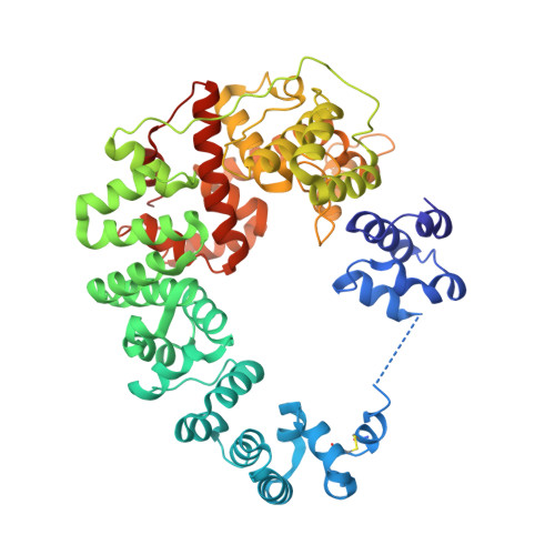
Crystal structure of soluble lytic transglycosylase Cj0843 of Campylobacter jejuni in complex with Bulgecin A


Experimental Data Snapshot

  • Method: X-RAY DIFFRACTION
  • Resolution: 2.04 Å
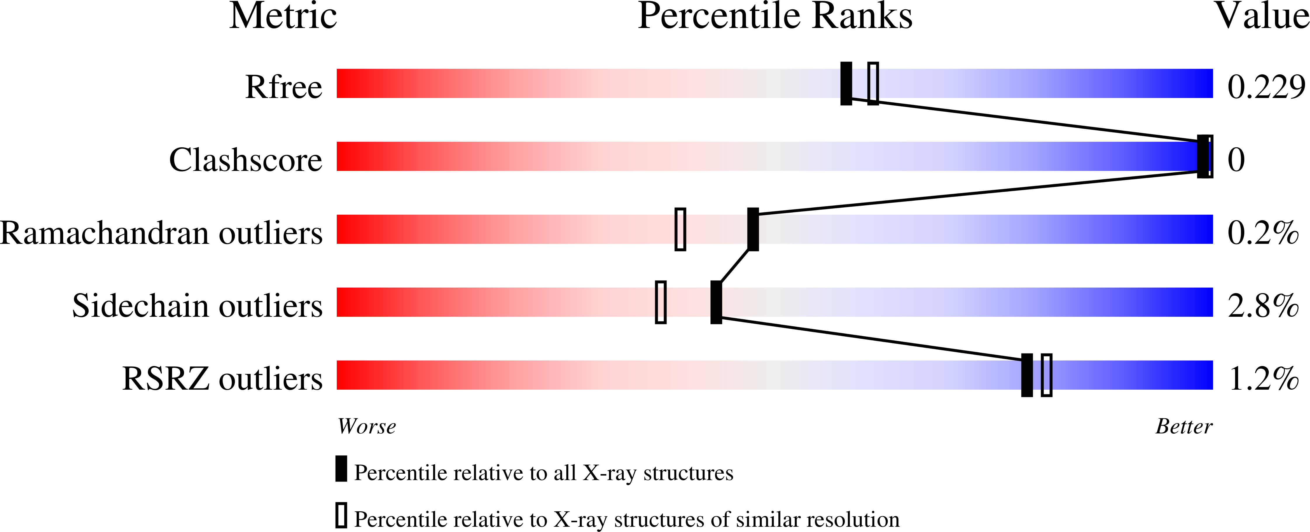
  • R-Value Free: 0.220 
  • R-Value Work: 0.177 
  • R-Value Observed: 0.179 

Starting Model: experimental
View more details

wwPDB Validation   3D Report Full Report


Ligand Structure Quality Assessment 


This is version 1.2 of the entry. See complete history


Literature

Structural studies and molecular dynamics simulations suggest a processive mechanism of exolytic lytic transglycosylase from Campylobacter jejuni.

Vijayaraghavan, J.Kumar, V.Krishnan, N.P.Kaufhold, R.T.Zeng, X.Lin, J.van den Akker, F.

(2018) PLoS One 13: e0197136-e0197136

  • DOI: https://doi.org/10.1371/journal.pone.0197136
  • Primary Citation of Related Structures:  
    6CF8, 6CF9, 6CFC

  • PubMed Abstract: 

    The bacterial soluble lytic transglycosylase (LT) breaks down the peptidoglycan (PG) layer during processes such as cell division. We present here crystal structures of the soluble LT Cj0843 from Campylobacter jejuni with and without bulgecin A inhibitor in the active site. Cj0843 has a doughnut shape similar but not identical to that of E. coli SLT70. The C-terminal catalytic domain is preceded by an L-domain, a large helical U-domain, a flexible linker, and a small N-terminal NU-domain. The flexible linker allows the NU-domain to reach over and complete the circular shape, using residues conserved in the Epsilonproteobacteria LT family. The inner surface of the Cj0843 doughnut is mostly positively charged including a pocket that has 8 Arg/Lys residues. Molecular dynamics simulations with PG strands revealed a potential functional role for this pocket in anchoring the negatively charged terminal tetrapeptide of the PG during several steps in the reaction including homing and aligning the PG strand for exolytic cleavage, and subsequent ratcheting of the PG strand to enhance processivity in degrading PG strands.


  • Organizational Affiliation

    Department of Biochemistry, Case Western Reserve University, Cleveland, OH, United States of America.


Macromolecules
Find similar proteins by:  (by identity cutoff)  |  3D Structure
Entity ID: 1
MoleculeChains Sequence LengthOrganismDetailsImage
Lytic transglycosylase524Campylobacter jejuniMutation(s): 0 
Gene Names: BD28_04025
Entity Groups  
Sequence Clusters30% Identity50% Identity70% Identity90% Identity95% Identity100% Identity
Sequence Annotations
Expand
  • Reference Sequence
Small Molecules
Ligands 2 Unique
IDChains Name / Formula / InChI Key2D Diagram3D Interactions
BLG (Subject of Investigation/LOI)
Query on BLG

Download Ideal Coordinates CCD File 
B [auth A]4-O-(4-O-SULFONYL-N-ACETYLGLUCOSAMININYL)-5-METHYLHYDROXY-L-PROLINE-TAURINE
C16 H30 N3 O14 S2
RPNZWZDLNYCCIG-HMMVDTEZSA-O
SO4
Query on SO4

Download Ideal Coordinates CCD File 
C [auth A]
D [auth A]
E [auth A]
F [auth A]
G [auth A]
C [auth A],
D [auth A],
E [auth A],
F [auth A],
G [auth A],
H [auth A],
I [auth A],
J [auth A],
K [auth A]
SULFATE ION
O4 S
QAOWNCQODCNURD-UHFFFAOYSA-L
Experimental Data & Validation

Experimental Data

  • Method: X-RAY DIFFRACTION
  • Resolution: 2.04 Å
  • R-Value Free: 0.220 
  • R-Value Work: 0.177 
  • R-Value Observed: 0.179 
  • Space Group: P 31 2 1
Unit Cell:
Length ( Å )Angle ( ˚ )
a = 74.294α = 90
b = 74.294β = 90
c = 190.461γ = 120
Software Package:
Software NamePurpose
XDSdata reduction
REFMACrefinement
PDB_EXTRACTdata extraction
XDSdata scaling
MOLREPphasing

Structure Validation

View Full Validation Report


Ligand Structure Quality Assessment 


Entry History 

Deposition Data

Revision History  (Full details and data files)

  • Version 1.0: 2018-05-30
    Type: Initial release
  • Version 1.1: 2018-06-06
    Changes: Data collection, Structure summary
  • Version 1.2: 2023-10-04
    Changes: Data collection, Database references, Refinement description