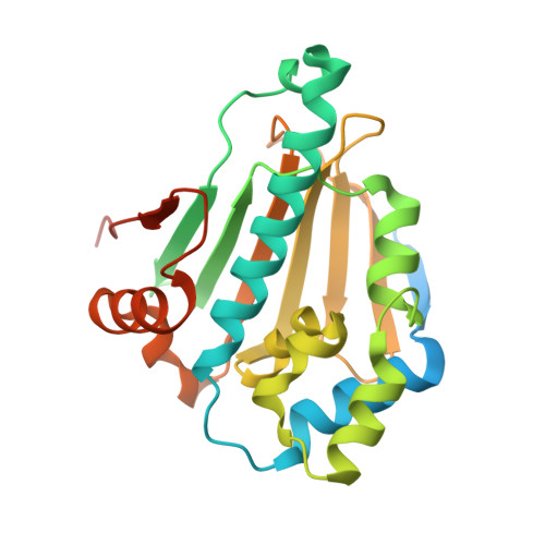
NECA derivatives exploit the paralog-specific properties of the site 3 side pocket of Grp94, the endoplasmic reticulum Hsp90.
Huck, J.D., Que, N.L.S., Immormino, R.M., Shrestha, L., Taldone, T., Chiosis, G., Gewirth, D.T.(2019) J Biol Chem 294: 16010-16019
- PubMed: 31501246
- DOI: https://doi.org/10.1074/jbc.RA119.009960
- Primary Citation of Related Structures:
6CYG, 6CYH, 6CYI, 6D1X - PubMed Abstract:
The hsp90 chaperones govern the function of essential client proteins critical for normal cell function as well as cancer initiation and progression. Hsp90 activity is driven by ATP, which binds to the N-terminal domain and induces large conformational changes that are required for client maturation. Inhibitors targeting the ATP-binding pocket of the N-terminal domain have anticancer effects, but most bind with similar affinity to cytosolic Hsp90α and Hsp90β, endoplasmic reticulum Grp94, and mitochondrial Trap1, the four cellular hsp90 paralogs. Paralog-specific inhibitors may lead to drugs with fewer side effects. The ATP-binding pockets of the four paralogs are flanked by three side pockets, termed sites 1, 2, and 3, which differ between the paralogs in their accessibility to inhibitors. Previous insights into the principles governing access to sites 1 and 2 have resulted in development of paralog-selective inhibitors targeting these sites, but the rules for selective targeting of site 3 are less clear. Earlier studies identified 5' N -ethylcarboxamido adenosine (NECA) as a Grp94-selective ligand. Here we use NECA and its derivatives to probe the properties of site 3. We found that derivatives that lengthen the 5' moiety of NECA improve selectivity for Grp94 over Hsp90α. Crystal structures reveal that the derivatives extend further into site 3 of Grp94 compared with their parent compound and that selectivity is due to paralog-specific differences in ligand pose and ligand-induced conformational strain in the protein. These studies provide a structural basis for Grp94-selective inhibition using site 3.
Organizational Affiliation:
Hauptman-Woodward Medical Research Institute, Buffalo, New York 14203.


