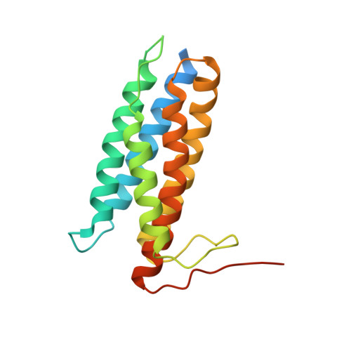
6D5K

Structure of Human ATP:Cobalamin Adenosyltransferase bound to ATP, and Adenosylcobalamin


Experimental Data Snapshot

  • Method: X-RAY DIFFRACTION
  • Resolution: 2.85 Å
  • R-Value Free: 0.239 
  • R-Value Work: 0.192 
  • R-Value Observed: 0.196 

Starting Model: experimental
View more details

wwPDB Validation   3D Report Full Report


Ligand Structure Quality Assessment 


This is version 1.4 of the entry. See complete history


Literature

Sacrificial Cobalt-Carbon Bond Homolysis in Coenzyme B12as a Cofactor Conservation Strategy.

Campanello, G.C.Ruetz, M.Dodge, G.J.Gouda, H.Gupta, A.Twahir, U.T.Killian, M.M.Watkins, D.Rosenblatt, D.S.Brunold, T.C.Warncke, K.Smith, J.L.Banerjee, R.

(2018) J Am Chem Soc 140: 13205-13208

  • DOI: https://doi.org/10.1021/jacs.8b08659
  • Primary Citation of Related Structures:  
    6D5K, 6D5X

  • PubMed Abstract: 

    A sophisticated intracellular trafficking pathway in humans is used to tailor vitamin B 12 into its active cofactor forms, and to deliver it to two known B 12 -dependent enzymes. Herein, we report an unexpected strategy for cellular retention of B 12 , an essential and reactive cofactor. If methylmalonyl-CoA mutase is unavailable to accept the coenzyme B 12 product of adenosyltransferase, the latter catalyzes homolytic scission of the cobalt-carbon bond in an unconventional reversal of the nucleophilic displacement reaction that was used to make it. The resulting homolysis product binds more tightly to adenosyltransferase than does coenzyme B 12 , facilitating cofactor retention. We have trapped, and characterized spectroscopically, an intermediate in which the cobalt-carbon bond is weakened prior to being broken. The physiological relevance of this sacrificial catalytic activity for cofactor retention is supported by the significantly lower coenzyme B 12 concentration in patients with dysfunctional methylmalonyl-CoA mutase but normal adenosyltransferase activity.


  • Organizational Affiliation

    Department of Biological Chemistry , University of Michigan , Ann Arbor , Michigan 48109-0600 , United States.


Macromolecules
Find similar proteins by:  (by identity cutoff)  |  3D Structure
Entity ID: 1
MoleculeChains Sequence LengthOrganismDetailsImage
Cob(I)yrinic acid a,c-diamide adenosyltransferase, mitochondrial
A, B, C
196Homo sapiensMutation(s): 0 
Gene Names: MMAB
EC: 2.5.1.17 (PDB Primary Data), 2.5.1 (UniProt)
UniProt & NIH Common Fund Data Resources
Find proteins for Q96EY8 (Homo sapiens)
Explore Q96EY8 
Go to UniProtKB:  Q96EY8
PHAROS:  Q96EY8
GTEx:  ENSG00000139428 
Entity Groups  
Sequence Clusters30% Identity50% Identity70% Identity90% Identity95% Identity100% Identity
UniProt GroupQ96EY8
Sequence Annotations
Expand
  • Reference Sequence
Small Molecules
Ligands 6 Unique
IDChains Name / Formula / InChI Key2D Diagram3D Interactions
B12 (Subject of Investigation/LOI)
Query on B12

Download Ideal Coordinates CCD File 
E [auth A],
F [auth A]
COBALAMIN
C62 H89 Co N13 O14 P
LKVIQTCSMMVGFU-DWSMJLPVSA-N
ATP (Subject of Investigation/LOI)
Query on ATP

Download Ideal Coordinates CCD File 
L [auth C]ADENOSINE-5'-TRIPHOSPHATE
C10 H16 N5 O13 P3
ZKHQWZAMYRWXGA-KQYNXXCUSA-N
5AD (Subject of Investigation/LOI)
Query on 5AD

Download Ideal Coordinates CCD File 
D [auth A],
I [auth B]
5'-DEOXYADENOSINE
C10 H13 N5 O3
XGYIMTFOTBMPFP-KQYNXXCUSA-N
EPE
Query on EPE

Download Ideal Coordinates CCD File 
N [auth C]4-(2-HYDROXYETHYL)-1-PIPERAZINE ETHANESULFONIC ACID
C8 H18 N2 O4 S
JKMHFZQWWAIEOD-UHFFFAOYSA-N
SO4
Query on SO4

Download Ideal Coordinates CCD File 
G [auth A]
H [auth A]
J [auth B]
K [auth B]
O [auth C]
G [auth A],
H [auth A],
J [auth B],
K [auth B],
O [auth C],
P [auth C]
SULFATE ION
O4 S
QAOWNCQODCNURD-UHFFFAOYSA-L
MG (Subject of Investigation/LOI)
Query on MG

Download Ideal Coordinates CCD File 
M [auth C],
Q [auth C]
MAGNESIUM ION
Mg
JLVVSXFLKOJNIY-UHFFFAOYSA-N
Experimental Data & Validation

Experimental Data

  • Method: X-RAY DIFFRACTION
  • Resolution: 2.85 Å
  • R-Value Free: 0.239 
  • R-Value Work: 0.192 
  • R-Value Observed: 0.196 
  • Space Group: P 31 2 1
Unit Cell:
Length ( Å )Angle ( ˚ )
a = 112.252α = 90
b = 112.252β = 90
c = 117.993γ = 120
Software Package:
Software NamePurpose
PHENIXrefinement
XDSdata reduction
Aimlessdata scaling
PHASERphasing

Structure Validation

View Full Validation Report


Ligand Structure Quality Assessment 


Entry History & Funding Information

Deposition Data


Funding OrganizationLocationGrant Number
National Institutes of Health/National Institute of Diabetes and Digestive and Kidney Disease (NIH/NIDDK)United StatesDK45776

Revision History  (Full details and data files)

  • Version 1.0: 2018-10-10
    Type: Initial release
  • Version 1.1: 2018-10-17
    Changes: Data collection, Database references, Structure summary
  • Version 1.2: 2018-10-31
    Changes: Data collection, Database references
  • Version 1.3: 2019-12-25
    Changes: Author supporting evidence
  • Version 1.4: 2023-10-04
    Changes: Data collection, Database references, Derived calculations, Refinement description