6E3W

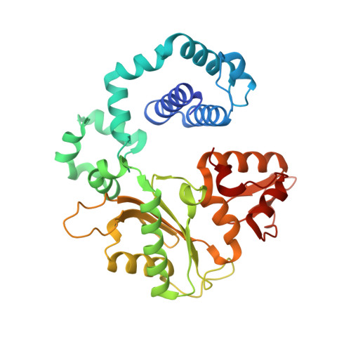
Structure of human DNA polymerase beta complexed with 8OA in the template base paired with incoming non-hydrolyzable GTP


Experimental Data Snapshot

  • Method: X-RAY DIFFRACTION
  • Resolution: 2.02 Å
  • R-Value Free: 0.245 
  • R-Value Work: 0.193 
  • R-Value Observed: 0.195 

Starting Models: experimental
View more details

wwPDB Validation   3D Report Full Report


Ligand Structure Quality Assessment 


This is version 1.2 of the entry. See complete history


Literature

Mutagenic Replication of the Major Oxidative Adenine Lesion 7,8-Dihydro-8-oxoadenine by Human DNA Polymerases.

Koag, M.C.Jung, H.Lee, S.

(2019) J Am Chem Soc 141: 4584-4596

  • DOI: https://doi.org/10.1021/jacs.8b08551
  • Primary Citation of Related Structures:  
    6E3R, 6E3V, 6E3W

  • PubMed Abstract: 

    Reactive oxygen species attack DNA to produce 7,8-dihyro-8-oxoguanine (oxoG) and 7,8-dihydro-8-oxoadenine (oxoA) as major lesions. The structural basis for the mutagenicity of oxoG, which induces G to T mutations, is well understood. However, the structural basis for the mutagenic potential of oxoA, which induces A to C mutations, remains poorly understood. To gain insight into oxoA-induced mutagenesis, we conducted kinetic studies of human DNA polymerases β and η replicating across oxoA and structural studies of polβ incorporating dTTP/dGTP opposite oxoA. While polη readily bypassed oxoA, it incorporated dGTP opposite oxoA with a catalytic specificity comparable to that of correct insertion, underscoring the promutagenic nature of the major oxidative adenine lesion. Polη and polβ incorporated dGTP opposite oxoA ∼170-fold and ∼100-fold more efficiently than that opposite dA, respectively, indicating that the 8-oxo moiety greatly facilitated error-prone replication. Crystal structures of polβ showed that, when paired with an incoming dTTP, the templating oxoA adopted an anti conformation and formed Watson-Crick base pair. When paired with dGTP, oxoA adopted a syn conformation and formed a Hoogsteen base pair with Watson-Crick-like geometry, highlighting the dual-coding potential of oxoA. The templating oxoA was stabilized by Lys280-mediated stacking and hydrogen bonds. Overall, these results provide insight into the mutagenic potential and dual-coding nature of the major oxidative adenine lesion.


  • Organizational Affiliation

    The Division of Chemical Biology and Medicinal Chemistry, College of Pharmacy , The University of Texas at Austin , Austin , Texas 78712 , United States.


Macromolecules

Find similar proteins by:  (by identity cutoff)  |  3D Structure
Entity ID: 1
MoleculeChains Sequence LengthOrganismDetailsImage
DNA polymerase beta335Homo sapiensMutation(s): 0 
Gene Names: POLB
EC: 2.7.7.7 (PDB Primary Data), 4.2.99 (PDB Primary Data), 4.2.99.18 (UniProt)
UniProt & NIH Common Fund Data Resources
Find proteins for P06746 (Homo sapiens)
Explore P06746 
Go to UniProtKB:  P06746
PHAROS:  P06746
GTEx:  ENSG00000070501 
Entity Groups  
Sequence Clusters30% Identity50% Identity70% Identity90% Identity95% Identity100% Identity
UniProt GroupP06746
Sequence Annotations
Expand
  • Reference Sequence

Find similar nucleic acids by:  Sequence   |   3D Structure  

Entity ID: 2
MoleculeChains LengthOrganismImage
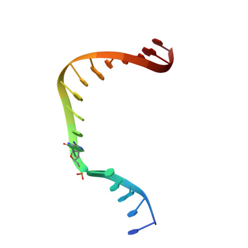
DNA (5'-D(*CP*CP*GP*AP*CP*(A38) P*TP*CP*GP*CP*AP*TP*CP*AP*GP*C)-3')B [auth T]16Homo sapiens
Sequence Annotations
Expand
  • Reference Sequence

Find similar nucleic acids by:  Sequence   |   3D Structure  

Entity ID: 3
MoleculeChains LengthOrganismImage
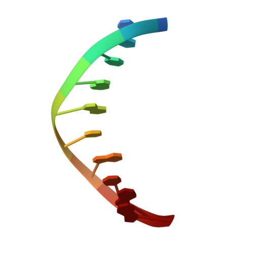
DNA (5'-D(*GP*CP*TP*GP*AP*TP*GP*CP*GP*A)-3')C [auth P]10Homo sapiens
Sequence Annotations
Expand
  • Reference Sequence

Find similar nucleic acids by:  Sequence   |   3D Structure  

Entity ID: 4
MoleculeChains LengthOrganismImage
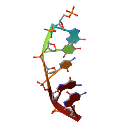
DNA (5'-D(P*GP*TP*CP*GP*G)-3')5Homo sapiens
Sequence Annotations
Expand
  • Reference Sequence
Experimental Data & Validation

Experimental Data

  • Method: X-RAY DIFFRACTION
  • Resolution: 2.02 Å
  • R-Value Free: 0.245 
  • R-Value Work: 0.193 
  • R-Value Observed: 0.195 
  • Space Group: P 1 21 1
Unit Cell:
Length ( Å )Angle ( ˚ )
a = 50.588α = 90
b = 79.594β = 107.54
c = 55.718γ = 90
Software Package:
Software NamePurpose
DENZOdata reduction
HKL-2000data scaling
MOLREPphasing
PHASERphasing
PHENIXrefinement

Structure Validation

View Full Validation Report


Ligand Structure Quality Assessment 


Entry History & Funding Information

Deposition Data


Funding OrganizationLocationGrant Number
National Institutes of Health/National Human Genome Research Institute (NIH/NHGRI)United StatesES-26676

Revision History  (Full details and data files)

  • Version 1.0: 2019-04-03
    Type: Initial release
  • Version 1.1: 2019-12-18
    Changes: Author supporting evidence
  • Version 1.2: 2023-10-11
    Changes: Data collection, Database references, Derived calculations, Refinement description