6E8S | pdb_00006e8s

Structure of the iMango-III aptamer bound to TO1-Biotin


Experimental Data Snapshot

  • Method: X-RAY DIFFRACTION
  • Resolution: 2.35 Å
  • R-Value Free: 
    0.207 (Depositor), 0.210 (DCC) 
  • R-Value Work: 
    0.182 (Depositor), 0.180 (DCC) 
  • R-Value Observed: 
    0.185 (Depositor) 

wwPDB Validation   3D Report Full Report


Ligand Structure Quality Assessment 

Created with Raphaël 2.3.0Worse 01 BetterLigand structure goodness of fit to experimental dataBest fitted EKJClick on this verticalbar to view detailsBest fitted SPMClick on this verticalbar to view details

This is version 1.3 of the entry. See complete history


Literature

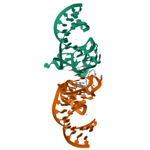
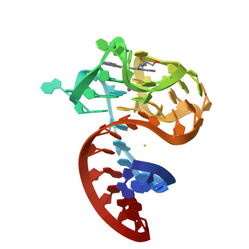
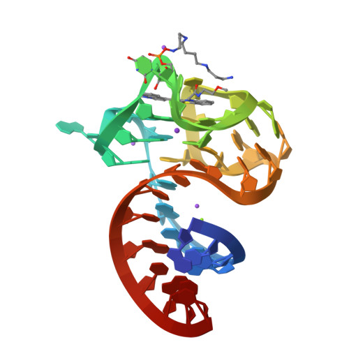
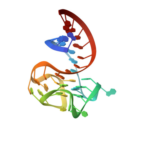
Structure and functional reselection of the Mango-III fluorogenic RNA aptamer.

Trachman 3rd., R.J.Autour, A.Jeng, S.C.Y.Abdolahzadeh, A.Andreoni, A.Cojocaru, R.Garipov, R.Dolgosheina, E.V.Knutson, J.R.Ryckelynck, M.Unrau, P.J.Ferre-D'Amare, A.R.

(2019) Nat Chem Biol 15: 472-479

  • DOI: https://doi.org/10.1038/s41589-019-0267-9
  • Primary Citation of Related Structures:  
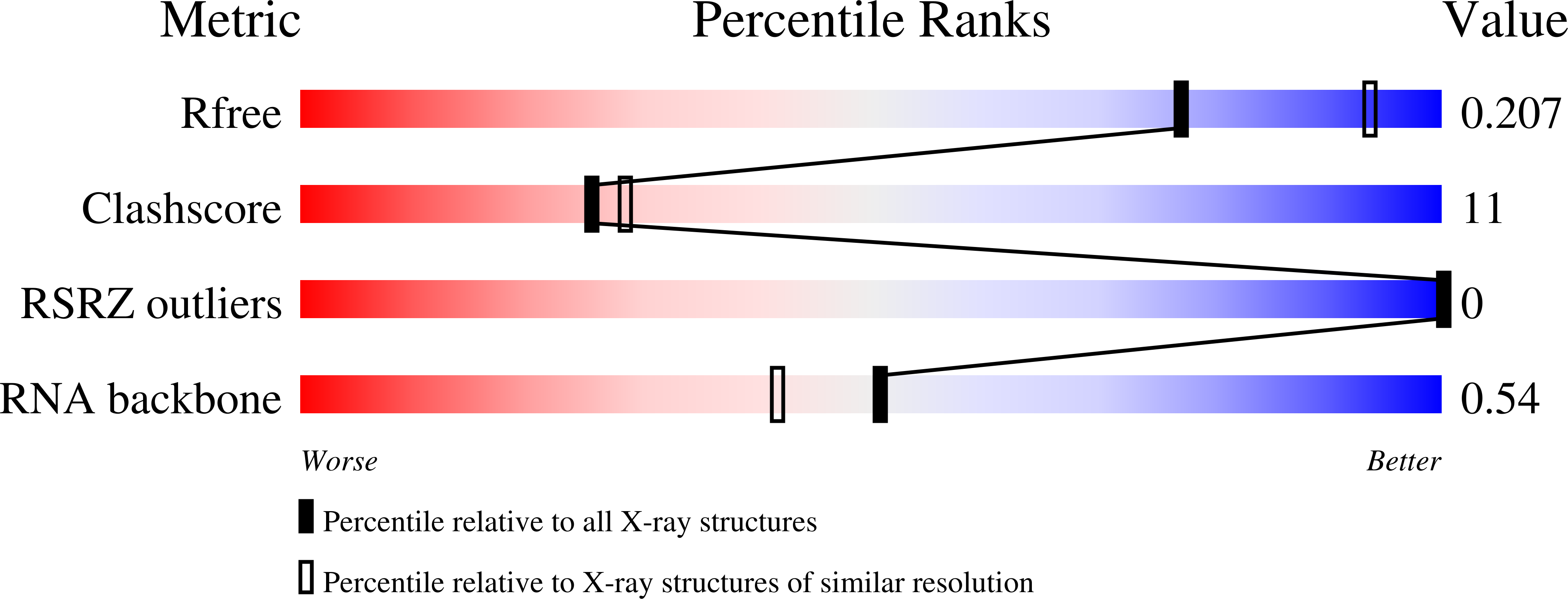
    6E8S, 6E8T, 6E8U

  • PubMed Abstract: 

    Several turn-on RNA aptamers that activate small-molecule fluorophores have been selected in vitro. Among these, the ~30 nucleotide Mango-III is notable because it binds the thiazole orange derivative TO1-Biotin with high affinity and fluoresces brightly (quantum yield 0.55). Uniquely among related aptamers, Mango-III exhibits biphasic thermal melting, characteristic of molecules with tertiary structure. We report crystal structures of TO1-Biotin complexes of Mango-III, a structure-guided mutant Mango-III(A10U), and a functionally reselected mutant iMango-III. The structures reveal a globular architecture arising from an unprecedented pseudoknot-like connectivity between a G-quadruplex and an embedded non-canonical duplex. The fluorophore is restrained into a planar conformation by the G-quadruplex, a lone, long-range trans Watson-Crick pair (whose A10U mutation increases quantum yield to 0.66), and a pyrimidine perpendicular to the nucleobase planes of those motifs. The improved iMango-III and Mango-III(A10U) fluoresce ~50% brighter than enhanced green fluorescent protein, making them suitable tags for live cell RNA visualization.


  • Organizational Affiliation

    Biochemistry and Biophysics Center, National Heart, Lung, and Blood Institute, Bethesda, MD, USA.


Macromolecules
Find similar nucleic acids by:  (by identity cutoff)  |  3D Structure
Entity ID: 1
MoleculeChains LengthOrganismImage
iMango-III aptamer
A, B
38synthetic construct
Sequence Annotations
Expand
  • Reference Sequence
Small Molecules
Ligands 5 Unique
IDChains Name / Formula / InChI Key2D Diagram3D Interactions
EKJ
Query on EKJ

Download Ideal Coordinates CCD File 
C [auth A],
G [auth B]
4-[(3-{2-[(2-methoxyethyl)amino]-2-oxoethyl}-1,3-benzothiazol-3-ium-2-yl)methyl]-1-methylquinolin-1-ium
C23 H25 N3 O2 S
WEFGJTSJMBBARG-UHFFFAOYSA-O
SPM
Query on SPM

Download Ideal Coordinates CCD File 
M [auth B]SPERMINE
C10 H26 N4
PFNFFQXMRSDOHW-UHFFFAOYSA-N
K
Query on K

Download Ideal Coordinates CCD File 
D [auth A],
I [auth B]
POTASSIUM ION
K
NPYPAHLBTDXSSS-UHFFFAOYSA-N
MG
Query on MG

Download Ideal Coordinates CCD File 
E [auth A],
J [auth B]
MAGNESIUM ION
Mg
JLVVSXFLKOJNIY-UHFFFAOYSA-N
NA
Query on NA

Download Ideal Coordinates CCD File 
F [auth A],
H [auth B],
K [auth B],
L [auth B]
SODIUM ION
Na
FKNQFGJONOIPTF-UHFFFAOYSA-N
Experimental Data & Validation

Experimental Data

  • Method: X-RAY DIFFRACTION
  • Resolution: 2.35 Å
  • R-Value Free:  0.207 (Depositor), 0.210 (DCC) 
  • R-Value Work:  0.182 (Depositor), 0.180 (DCC) 
  • R-Value Observed: 0.185 (Depositor) 
Space Group: P 21 21 21
Unit Cell:
Length ( Å )Angle ( ˚ )
a = 63.031α = 90
b = 67.585β = 90
c = 76.667γ = 90
Software Package:
Software NamePurpose
PHENIXrefinement
XDSdata reduction
Aimlessdata scaling
SHELXDphasing

Structure Validation

View Full Validation Report


Ligand Structure Quality Assessment 

Created with Raphaël 2.3.0Worse 01 BetterLigand structure goodness of fit to experimental dataBest fitted EKJClick on this verticalbar to view detailsBest fitted SPMClick on this verticalbar to view details

Entry History 

Deposition Data

Revision History  (Full details and data files)

  • Version 1.0: 2019-04-17
    Type: Initial release
  • Version 1.1: 2019-04-24
    Changes: Advisory, Data collection, Database references, Derived calculations
  • Version 1.2: 2019-05-15
    Changes: Data collection, Structure summary
  • Version 1.3: 2024-03-13
    Changes: Data collection, Database references, Derived calculations