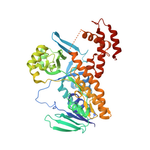
6FOY

The crystal structure of P.fluorescens Kynurenine 3-monooxygenase (KMO) in complex with competitive inhibitor No. 9

  • Classification: OXIDOREDUCTASE
  • Organism(s): Pseudomonas fluorescens
  • Expression System: Escherichia coli
  • Mutation(s): Yes 

  • Deposited: 2018-02-08 Released: 2019-08-21 
  • Deposition Author(s): Levy, C.W., Leys, D.
  • Funding Organization(s): Biotechnology and Biological Sciences Research Council, Engineering and Physical Sciences Research Council, Medical Research Council (United Kingdom)

Experimental Data Snapshot

  • Method: X-RAY DIFFRACTION
  • Resolution: 1.65 Å
  • R-Value Free: 0.213 
  • R-Value Work: 0.164 
  • R-Value Observed: 0.165 

Starting Model: other
View more details

wwPDB Validation   3D Report Full Report


Ligand Structure Quality Assessment 


This is version 1.2 of the entry. See complete history


Literature

A brain-permeable inhibitor of the neurodegenerative disease target kynurenine 3-monooxygenase prevents accumulation of neurotoxic metabolites.

Zhang, S.Sakuma, M.Deora, G.S.Levy, C.W.Klausing, A.Breda, C.Read, K.D.Edlin, C.D.Ross, B.P.Wright Muelas, M.Day, P.J.O'Hagan, S.Kell, D.B.Schwarcz, R.Leys, D.Heyes, D.J.Giorgini, F.Scrutton, N.S.

(2019) Commun Biol 2: 271-271

  • DOI: https://doi.org/10.1038/s42003-019-0520-5
  • Primary Citation of Related Structures:  
    6FOX, 6FOY, 6FOZ, 6FP0, 6FP1, 6FPH

  • PubMed Abstract: 

    Dysregulation of the kynurenine pathway (KP) leads to imbalances in neuroactive metabolites associated with the pathogenesis of several neurodegenerative disorders, including Huntington's disease (HD). Inhibition of the enzyme kynurenine 3-monooxygenase (KMO) in the KP normalises these metabolic imbalances and ameliorates neurodegeneration and related phenotypes in several neurodegenerative disease models. KMO is thus a promising candidate drug target for these disorders, but known inhibitors are not brain permeable. Here, 19 new KMO inhibitors have been identified. One of these ( 1 ) is neuroprotective in a Drosophila HD model but is minimally brain penetrant in mice. The prodrug variant ( 1b ) crosses the blood-brain barrier, releases 1 in the brain, thereby lowering levels of 3-hydroxykynurenine, a toxic KP metabolite linked to neurodegeneration. Prodrug 1b will advance development of targeted therapies against multiple neurodegenerative and neuroinflammatory diseases in which KP likely plays a role, including HD, Alzheimer's disease, and Parkinson's disease.


  • Organizational Affiliation

    1Manchester Institute of Biotechnology and School of Chemistry, The University of Manchester, Manchester, M1 7DN UK.


Macromolecules
Find similar proteins by:  (by identity cutoff)  |  3D Structure
Entity ID: 1
MoleculeChains Sequence LengthOrganismDetailsImage
Kynurenine 3-monooxygenase
A, B
460Pseudomonas fluorescensMutation(s): 1 
Gene Names: kmoqbsG
EC: 1.14.13.9
UniProt
Find proteins for Q84HF5 (Pseudomonas fluorescens)
Explore Q84HF5 
Go to UniProtKB:  Q84HF5
Entity Groups  
Sequence Clusters30% Identity50% Identity70% Identity90% Identity95% Identity100% Identity
UniProt GroupQ84HF5
Sequence Annotations
Expand
  • Reference Sequence
Small Molecules
Ligands 4 Unique
IDChains Name / Formula / InChI Key2D Diagram3D Interactions
FAD
Query on FAD

Download Ideal Coordinates CCD File 
C [auth A],
G [auth B]
FLAVIN-ADENINE DINUCLEOTIDE
C27 H33 N9 O15 P2
VWWQXMAJTJZDQX-UYBVJOGSSA-N
E0T (Subject of Investigation/LOI)
Query on E0T

Download Ideal Coordinates CCD File 
D [auth A],
H [auth B]
5-[2,3-bis(chloranyl)phenyl]furan-2-carboxylic acid
C11 H6 Cl2 O3
BSMKDXIJGSTBNT-UHFFFAOYSA-N
PGE
Query on PGE

Download Ideal Coordinates CCD File 
E [auth A],
I [auth B]
TRIETHYLENE GLYCOL
C6 H14 O4
ZIBGPFATKBEMQZ-UHFFFAOYSA-N
CA
Query on CA

Download Ideal Coordinates CCD File 
F [auth A]CALCIUM ION
Ca
BHPQYMZQTOCNFJ-UHFFFAOYSA-N
Experimental Data & Validation

Experimental Data

  • Method: X-RAY DIFFRACTION
  • Resolution: 1.65 Å
  • R-Value Free: 0.213 
  • R-Value Work: 0.164 
  • R-Value Observed: 0.165 
  • Space Group: P 1 21 1
Unit Cell:
Length ( Å )Angle ( ˚ )
a = 69.8α = 90
b = 52.977β = 103.833
c = 135.765γ = 90
Software Package:
Software NamePurpose
PHENIXrefinement
xia2data reduction
xia2data scaling
PHASERphasing

Structure Validation

View Full Validation Report


Ligand Structure Quality Assessment 


Entry History & Funding Information

Deposition Data


Funding OrganizationLocationGrant Number
Biotechnology and Biological Sciences Research CouncilUnited KingdomBB/P009042/1
Biotechnology and Biological Sciences Research CouncilUnited KingdomBB/R000093/1
Engineering and Physical Sciences Research CouncilUnited KingdomEP/J020192/1
Medical Research Council (United Kingdom)United KingdomMR/N00373X/1

Revision History  (Full details and data files)

  • Version 1.0: 2019-08-21
    Type: Initial release
  • Version 1.1: 2020-03-04
    Changes: Database references
  • Version 1.2: 2024-05-01
    Changes: Data collection, Database references, Derived calculations, Refinement description