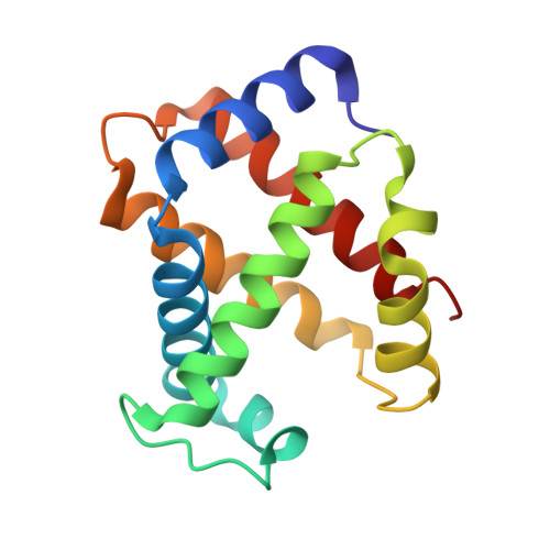
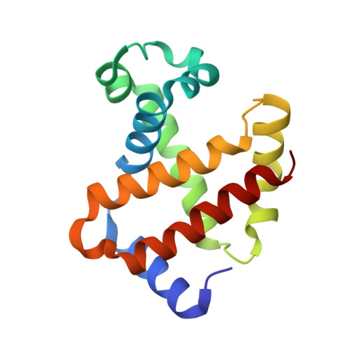
6IHX

Crystal Structure Analysis of bovine Hemoglobin modified by SNP


Experimental Data Snapshot

  • Method: X-RAY DIFFRACTION
  • Resolution: 1.46 Å
  • R-Value Free: 0.200 
  • R-Value Work: 0.145 
  • R-Value Observed: 0.146 

Starting Model: experimental
View more details

wwPDB Validation   3D Report Full Report


Ligand Structure Quality Assessment 


This is version 1.2 of the entry. See complete history


Literature

Quaternary Structure Analysis of a Hemoglobin Core in Hemoglobin-Albumin Cluster.

Morita, Y.Yamada, T.Kureishi, M.Kihira, K.Komatsu, T.

(2018) J Phys Chem B 122: 12031-12039

  • DOI: https://doi.org/10.1021/acs.jpcb.8b10077
  • Primary Citation of Related Structures:  
    6IHX, 6II1

  • PubMed Abstract: 

    A core-shell ensemble of bovine hemoglobin (Hb) and human serum albumin (HSA) is an artificial O 2 carrier as a red blood cell substitute. This protein particle is created by covalent wrapping of a carbonyl Hb with HSAs: Hb R -HSA 3 cluster, where Hb R signifies the use of carbonyl Hb (relaxed (R) state conformation) as a starting material. The Hb R -HSA 3 cluster exhibits high O 2 affinity and low cooperativity. Analysis of the quaternary structure of the central Hb R in the cluster revealed that its high O 2 affinity is attributed to the physically immobile Hb R nucleus. Circular dichroism and UV-vis absorption spectroscopy showed that the structure of deoxy Hb R core closely resembles the R-state. The crystal structure of Lys-modified carbonyl Hb R was superimposed on that of carbonyl Hb. These results imply that chemical modifications of the surface Lys groups and Cys-93(β) of the carbonyl Hb with cross-linking agent interfered in the quaternary structure movement from the R-state to tense (T) state. As expected, coupling of deoxy Hb (T-state) with HSAs yielded Hb T -HSA 3 cluster having low O 2 affinity. The mixing of Hb R -HSA 3 and Hb T -HSA 3 clusters conferred a tailor-made formulation of artificial O 2 carrier with a desired O 2 affinity ( P 50 ).


  • Organizational Affiliation

    Department of Applied Chemistry, Faculty of Science and Engineering , Chuo University , 1-13-27 Kasuga , Bunkyo-ku , Tokyo 112-8551 , Japan.


Macromolecules
Find similar proteins by:  (by identity cutoff)  |  3D Structure
Entity ID: 1
MoleculeChains Sequence LengthOrganismDetailsImage
Hemoglobin subunit alpha
A, C
140Bos taurusMutation(s): 0 
UniProt
Find proteins for P01966 (Bos taurus)
Explore P01966 
Go to UniProtKB:  P01966
Entity Groups  
Sequence Clusters30% Identity50% Identity70% Identity90% Identity95% Identity100% Identity
UniProt GroupP01966
Sequence Annotations
Expand
  • Reference Sequence
Find similar proteins by:  (by identity cutoff)  |  3D Structure
Entity ID: 2
MoleculeChains Sequence LengthOrganismDetailsImage
Hemoglobin subunit beta
B, D
144Bos taurusMutation(s): 0 
UniProt
Find proteins for P02070 (Bos taurus)
Explore P02070 
Go to UniProtKB:  P02070
Entity Groups  
Sequence Clusters30% Identity50% Identity70% Identity90% Identity95% Identity100% Identity
UniProt GroupP02070
Sequence Annotations
Expand
  • Reference Sequence
Small Molecules
Ligands 2 Unique
IDChains Name / Formula / InChI Key2D Diagram3D Interactions
HEM
Query on HEM

Download Ideal Coordinates CCD File 
E [auth A],
G [auth B],
I [auth C],
K [auth D]
PROTOPORPHYRIN IX CONTAINING FE
C34 H32 Fe N4 O4
KABFMIBPWCXCRK-RGGAHWMASA-L
CMO
Query on CMO

Download Ideal Coordinates CCD File 
F [auth A],
H [auth B],
J [auth C],
L [auth D]
CARBON MONOXIDE
C O
UGFAIRIUMAVXCW-UHFFFAOYSA-N
Experimental Data & Validation

Experimental Data

  • Method: X-RAY DIFFRACTION
  • Resolution: 1.46 Å
  • R-Value Free: 0.200 
  • R-Value Work: 0.145 
  • R-Value Observed: 0.146 
  • Space Group: P 21 21 21
Unit Cell:
Length ( Å )Angle ( ˚ )
a = 65.117α = 90
b = 78.132β = 90
c = 109.539γ = 90
Software Package:
Software NamePurpose
HKL-2000data scaling
REFMACrefinement
PDB_EXTRACTdata extraction
HKL-2000data reduction
PHASERphasing

Structure Validation

View Full Validation Report


Ligand Structure Quality Assessment 


Entry History 

Deposition Data

Revision History  (Full details and data files)

  • Version 1.0: 2018-12-12
    Type: Initial release
  • Version 1.1: 2019-04-24
    Changes: Data collection, Database references
  • Version 1.2: 2023-11-22
    Changes: Data collection, Database references, Refinement description