6KYL

Crystal Structure of Phosphatidic acid Transporter Ups1/Mdm35 in Complex with (2R)-3-(phosphonooxy)propane-1,2-diyl dihexanoate


Experimental Data Snapshot

  • Method: X-RAY DIFFRACTION
  • Resolution: 3.55 Å
  • R-Value Free: 0.310 
  • R-Value Work: 0.287 
  • R-Value Observed: 0.289 

Starting Model: experimental
View more details

wwPDB Validation   3D Report Full Report


Ligand Structure Quality Assessment 


This is version 1.4 of the entry. See complete history


Literature

Molecular mechanism of mitochondrial phosphatidate transfer by Ups1.

Lu, J.Chan, C.Yu, L.Fan, J.Sun, F.Zhai, Y.

(2020) Commun Biol 3: 468-468

  • DOI: https://doi.org/10.1038/s42003-020-01121-x
  • Primary Citation of Related Structures:  
    6KYL

  • PubMed Abstract: 

    Cardiolipin, an essential mitochondrial physiological regulator, is synthesized from phosphatidic acid (PA) in the inner mitochondrial membrane (IMM). PA is synthesized in the endoplasmic reticulum and transferred to the IMM via the outer mitochondrial membrane (OMM) under mediation by the Ups1/Mdm35 protein family. Despite the availability of numerous crystal structures, the detailed mechanism underlying PA transfer between mitochondrial membranes remains unclear. Here, a model of Ups1/Mdm35-membrane interaction is established using combined crystallographic data, all-atom molecular dynamics simulations, extensive structural comparisons, and biophysical assays. The α2-loop, L2-loop, and α3 helix of Ups1 mediate membrane interactions. Moreover, non-complexed Ups1 on membranes is found to be a key transition state for PA transfer. The membrane-bound non-complexed Ups1/ membrane-bound Ups1 ratio, which can be regulated by environmental pH, is inversely correlated with the PA transfer activity of Ups1/Mdm35. These results demonstrate a new model of the fine conformational changes of Ups1/Mdm35 during PA transfer.


  • Organizational Affiliation

    National Laboratory of Biomacromolecules, CAS Center for Excellence in Biomacromolecules, Institute of Biophysics, Chinese Academy of Sciences, 100101, Beijing, China.


Macromolecules
Find similar proteins by:  (by identity cutoff)  |  3D Structure
Entity ID: 1
MoleculeChains Sequence LengthOrganismDetailsImage
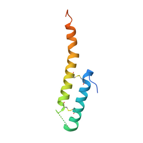
Mitochondrial distribution and morphology protein 35
A, C
86Saccharomyces cerevisiae S288CMutation(s): 0 
Gene Names: MDM35YKL053C-A
UniProt
Find proteins for O60200 (Saccharomyces cerevisiae (strain ATCC 204508 / S288c))
Explore O60200 
Go to UniProtKB:  O60200
Entity Groups  
Sequence Clusters30% Identity50% Identity70% Identity90% Identity95% Identity100% Identity
UniProt GroupO60200
Sequence Annotations
Expand
  • Reference Sequence
Find similar proteins by:  (by identity cutoff)  |  3D Structure
Entity ID: 2
MoleculeChains Sequence LengthOrganismDetailsImage
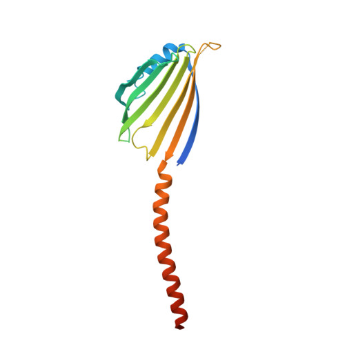
Protein UPS1, mitochondrial
B, D
189Saccharomyces cerevisiae S288CMutation(s): 0 
Gene Names: UPS1YLR193C
UniProt
Find proteins for Q05776 (Saccharomyces cerevisiae (strain ATCC 204508 / S288c))
Explore Q05776 
Go to UniProtKB:  Q05776
Entity Groups  
Sequence Clusters30% Identity50% Identity70% Identity90% Identity95% Identity100% Identity
UniProt GroupQ05776
Sequence Annotations
Expand
  • Reference Sequence
Small Molecules
Ligands 1 Unique
IDChains Name / Formula / InChI Key2D Diagram3D Interactions
44E (Subject of Investigation/LOI)
Query on 44E

Download Ideal Coordinates CCD File 
E [auth B],
F [auth D]
(2R)-3-(phosphonooxy)propane-1,2-diyl dihexanoate
C15 H29 O8 P
SFZZRGHNPILUOD-CYBMUJFWSA-N
Experimental Data & Validation

Experimental Data

  • Method: X-RAY DIFFRACTION
  • Resolution: 3.55 Å
  • R-Value Free: 0.310 
  • R-Value Work: 0.287 
  • R-Value Observed: 0.289 
  • Space Group: P 41
Unit Cell:
Length ( Å )Angle ( ˚ )
a = 90.14α = 90
b = 90.14β = 90
c = 81.319γ = 90
Software Package:
Software NamePurpose
PHENIXrefinement
SCALEPACKdata scaling
PDB_EXTRACTdata extraction
HKL-2000data reduction
PHASERphasing

Structure Validation

View Full Validation Report


Ligand Structure Quality Assessment 


Entry History 

Deposition Data

Revision History  (Full details and data files)

  • Version 1.0: 2019-10-02
    Type: Initial release
  • Version 1.1: 2020-09-02
    Changes: Database references
  • Version 1.2: 2020-09-09
    Changes: Database references
  • Version 1.3: 2023-11-22
    Changes: Data collection, Database references, Refinement description
  • Version 1.4: 2024-10-23
    Changes: Structure summary