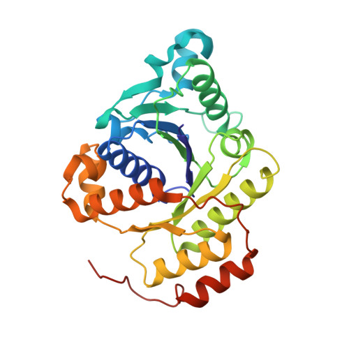
6N4M

IDS-oxidized ADP-bound form of the nitrogenase Fe-protein from A. vinelandii


Experimental Data Snapshot

  • Method: X-RAY DIFFRACTION
  • Resolution: 1.58 Å
  • R-Value Free: 0.188 
  • R-Value Work: 0.164 
  • R-Value Observed: 0.165 

Starting Model: experimental
View more details

wwPDB Validation   3D Report Full Report


Ligand Structure Quality Assessment 


This is version 1.3 of the entry. See complete history


Literature

Site-Specific Oxidation State Assignments of the Iron Atoms in the [4Fe:4S]2+/1+/0States of the Nitrogenase Fe-Protein.

Wenke, B.B.Spatzal, T.Rees, D.C.

(2019) Angew Chem Int Ed Engl 58: 3894-3897

  • DOI: https://doi.org/10.1002/anie.201813966
  • Primary Citation of Related Structures:  
    6N4J, 6N4K, 6N4L, 6N4M

  • PubMed Abstract: 

    The nitrogenase iron protein (Fe-protein) contains an unusual [4Fe:4S] iron-sulphur cluster that is stable in three oxidation states: 2+, 1+, and 0. Here, we use spatially resolved anomalous dispersion (SpReAD) refinement to determine oxidation assignments for the individual irons for each state. Additionally, we report the 1.13-Å resolution structure for the ADP bound Fe-protein, the highest resolution Fe-protein structure presently determined. In the dithionite-reduced [4Fe:4S] 1+ state, our analysis identifies a solvent exposed, delocalized Fe 2.5+ pair and a buried Fe 2+ pair. We propose that ATP binding by the Fe-protein promotes an internal redox rearrangement such that the solvent-exposed Fe pair becomes reduced, thereby facilitating electron transfer to the nitrogenase molybdenum iron-protein. In the [4Fe:4S] 0 and [4Fe:4S] 2+ states, the SpReAD analysis supports oxidation states assignments for all irons in these clusters of Fe 2+ and valence delocalized Fe 2.5+ , respectively.


  • Organizational Affiliation

    Division of Chemistry and Chemical Engineering, California Institute of Technology, Pasadena, CA, 91125, USA.


Macromolecules
Find similar proteins by:  (by identity cutoff)  |  3D Structure
Entity ID: 1
MoleculeChains Sequence LengthOrganismDetailsImage
Nitrogenase iron protein 1289Azotobacter vinelandiiMutation(s): 0 
EC: 1.18.6.1
UniProt
Find proteins for P00459 (Azotobacter vinelandii)
Explore P00459 
Go to UniProtKB:  P00459
Entity Groups  
Sequence Clusters30% Identity50% Identity70% Identity90% Identity95% Identity100% Identity
UniProt GroupP00459
Sequence Annotations
Expand
  • Reference Sequence
Experimental Data & Validation

Experimental Data

  • Method: X-RAY DIFFRACTION
  • Resolution: 1.58 Å
  • R-Value Free: 0.188 
  • R-Value Work: 0.164 
  • R-Value Observed: 0.165 
  • Space Group: P 2 21 21
Unit Cell:
Length ( Å )Angle ( ˚ )
a = 45.806α = 90
b = 74.579β = 90
c = 75.02γ = 90
Software Package:
Software NamePurpose
REFMACrefinement
XDSdata reduction
SCALAdata scaling
PHASERphasing
PDB_EXTRACTdata extraction

Structure Validation

View Full Validation Report


Ligand Structure Quality Assessment 


Entry History & Funding Information

Deposition Data


Funding OrganizationLocationGrant Number
National Institutes of Health/National Institute of General Medical Sciences (NIH/NIGMS)United StatesGM045162

Revision History  (Full details and data files)

  • Version 1.0: 2019-02-13
    Type: Initial release
  • Version 1.1: 2019-03-27
    Changes: Data collection, Database references
  • Version 1.2: 2020-01-01
    Changes: Author supporting evidence
  • Version 1.3: 2023-10-11
    Changes: Data collection, Database references, Refinement description