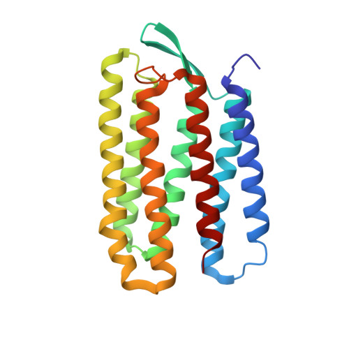
6RQO

Steady-state-SMX activated state structure of bacteriorhodopsin


Experimental Data Snapshot

  • Method: X-RAY DIFFRACTION
  • Resolution: 2.00 Å
  • R-Value Free: 0.246 
  • R-Value Work: 0.205 
  • R-Value Observed: 0.207 

Starting Model: experimental
View more details

wwPDB Validation   3D Report Full Report


Ligand Structure Quality Assessment 


This is version 1.2 of the entry. See complete history


Literature

Proton uptake mechanism in bacteriorhodopsin captured by serial synchrotron crystallography.

Weinert, T.Skopintsev, P.James, D.Dworkowski, F.Panepucci, E.Kekilli, D.Furrer, A.Brunle, S.Mous, S.Ozerov, D.Nogly, P.Wang, M.Standfuss, J.

(2019) Science 365: 61-65

  • DOI: https://doi.org/10.1126/science.aaw8634
  • Primary Citation of Related Structures:  
    6RNJ, 6RPH, 6RQO, 6RQP

  • PubMed Abstract: 

    Conformational dynamics are essential for proteins to function. We adapted time-resolved serial crystallography developed at x-ray lasers to visualize protein motions using synchrotrons. We recorded the structural changes in the light-driven proton-pump bacteriorhodopsin over 200 milliseconds in time. The snapshot from the first 5 milliseconds after photoactivation shows structural changes associated with proton release at a quality comparable to that of previous x-ray laser experiments. From 10 to 15 milliseconds onwards, we observe large additional structural rearrangements up to 9 angstroms on the cytoplasmic side. Rotation of leucine-93 and phenylalanine-219 opens a hydrophobic barrier, leading to the formation of a water chain connecting the intracellular aspartic acid-96 with the retinal Schiff base. The formation of this proton wire recharges the membrane pump with a proton for the next cycle.


  • Organizational Affiliation

    Division of Biology and Chemistry-Laboratory for Biomolecular Research, Paul Scherrer Institut, 5232 Villigen, Switzerland. tobias.weinert@psi.ch.


Macromolecules
Find similar proteins by:  (by identity cutoff)  |  3D Structure
Entity ID: 1
MoleculeChains Sequence LengthOrganismDetailsImage
Bacteriorhodopsin223Halobacterium salinarum NRC-1Mutation(s): 0 
Membrane Entity: Yes 
UniProt
Find proteins for P02945 (Halobacterium salinarum (strain ATCC 700922 / JCM 11081 / NRC-1))
Explore P02945 
Go to UniProtKB:  P02945
Entity Groups  
Sequence Clusters30% Identity50% Identity70% Identity90% Identity95% Identity100% Identity
UniProt GroupP02945
Sequence Annotations
Expand
  • Reference Sequence
Small Molecules
Experimental Data & Validation

Experimental Data

  • Method: X-RAY DIFFRACTION
  • Resolution: 2.00 Å
  • R-Value Free: 0.246 
  • R-Value Work: 0.205 
  • R-Value Observed: 0.207 
  • Space Group: P 63
Unit Cell:
Length ( Å )Angle ( ˚ )
a = 62α = 90
b = 62β = 90
c = 110.3γ = 120
Software Package:
Software NamePurpose
PHENIXrefinement
PDB_EXTRACTdata extraction
CrystFELdata reduction
CrystFELdata scaling
PHASERphasing

Structure Validation

View Full Validation Report


Ligand Structure Quality Assessment 


Entry History 

Deposition Data

Revision History  (Full details and data files)

  • Version 1.0: 2019-07-17
    Type: Initial release
  • Version 1.1: 2019-08-21
    Changes: Data collection
  • Version 1.2: 2024-01-24
    Changes: Advisory, Data collection, Database references, Refinement description