6T7F

RCR E3 ligase E2-Ubiquitin transthiolation intermediate


Experimental Data Snapshot

  • Method: X-RAY DIFFRACTION
  • Resolution: 2.58 Å
  • R-Value Free: 
    0.256 (Depositor), 0.260 (DCC) 
  • R-Value Work: 
    0.195 (Depositor), 0.200 (DCC) 
  • R-Value Observed: 
    0.198 (Depositor) 

Starting Models: experimental
View more details

wwPDB Validation   3D Report Full Report


Ligand Structure Quality Assessment 

Created with Raphaël 2.3.0Worse 01 BetterLigand structure goodness of fit to experimental dataBest fitted LWZClick on this verticalbar to view details

This is version 1.4 of the entry. See complete history


Literature

Structural basis for RING-Cys-Relay E3 ligase activity and its role in axon integrity.

Mabbitt, P.D.Loreto, A.Dery, M.A.Fletcher, A.J.Stanley, M.Pao, K.C.Wood, N.T.Coleman, M.P.Virdee, S.

(2020) Nat Chem Biol 16: 1227-1236

  • DOI: https://doi.org/10.1038/s41589-020-0598-6
  • Primary Citation of Related Structures:  
    6T7F

  • PubMed Abstract: 

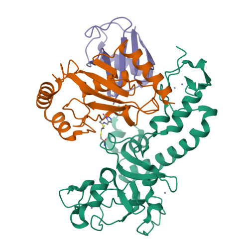
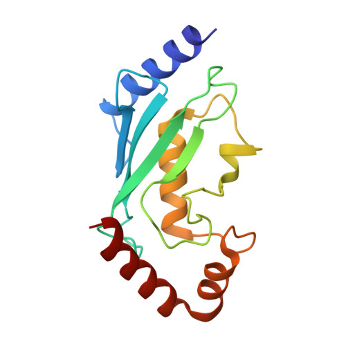
    MYCBP2 is a ubiquitin (Ub) E3 ligase (E3) that is essential for neurodevelopment and regulates axon maintenance. MYCBP2 transfers Ub to nonlysine substrates via a newly discovered RING-Cys-Relay (RCR) mechanism, where Ub is relayed from an upstream cysteine to a downstream substrate esterification site. The molecular bases for E2-E3 Ub transfer and Ub relay are unknown. Whether these activities are linked to the neural phenotypes is also unclear. We describe the crystal structure of a covalently trapped E2~Ub:MYCBP2 transfer intermediate revealing key structural rearrangements upon E2-E3 Ub transfer and Ub relay. Our data suggest that transfer to the dynamic upstream cysteine, whilst mitigating lysine activity, requires a closed-like E2~Ub conjugate with tempered reactivity, and Ub relay is facilitated by a helix-coil transition. Furthermore, neurodevelopmental defects and delayed injury-induced degeneration in RCR-defective knock-in mice suggest its requirement, and that of substrate esterification activity, for normal neural development and programmed axon degeneration.


  • Organizational Affiliation

    MRC Protein Phosphorylation and Ubiquitylation Unit, University of Dundee, Dundee, UK.


Macromolecules
Find similar proteins by:  (by identity cutoff)  |  3D Structure
Entity ID: 1
MoleculeChains Sequence LengthOrganismDetailsImage
E3 ubiquitin-protein ligase MYCBP2261Homo sapiensMutation(s): 0 
Gene Names: MYCBP2KIAA0916PAM
EC: 2.3.2 (PDB Primary Data), 2.3.2.33 (UniProt)
UniProt & NIH Common Fund Data Resources
Find proteins for O75592 (Homo sapiens)
Explore O75592 
Go to UniProtKB:  O75592
PHAROS:  O75592
GTEx:  ENSG00000005810 
Entity Groups  
Sequence Clusters30% Identity50% Identity70% Identity90% Identity95% Identity100% Identity
UniProt GroupO75592
Sequence Annotations
Expand
  • Reference Sequence
Find similar proteins by:  (by identity cutoff)  |  3D Structure
Entity ID: 2
MoleculeChains Sequence LengthOrganismDetailsImage
Ubiquitin-conjugating enzyme E2 D3150Homo sapiensMutation(s): 4 
Gene Names: UBE2D3UBC5CUBCH5C
EC: 2.3.2.23 (PDB Primary Data), 2.3.2.24 (PDB Primary Data)
UniProt & NIH Common Fund Data Resources
Find proteins for P61077 (Homo sapiens)
Explore P61077 
Go to UniProtKB:  P61077
PHAROS:  P61077
GTEx:  ENSG00000109332 
Entity Groups  
Sequence Clusters30% Identity50% Identity70% Identity90% Identity95% Identity100% Identity
UniProt GroupP61077
Sequence Annotations
Expand
  • Reference Sequence
Find similar proteins by:  (by identity cutoff)  |  3D Structure
Entity ID: 3
MoleculeChains Sequence LengthOrganismDetailsImage
Polyubiquitin-C73Homo sapiensMutation(s): 0 
Gene Names: UBC
UniProt & NIH Common Fund Data Resources
Find proteins for P0CG48 (Homo sapiens)
Explore P0CG48 
Go to UniProtKB:  P0CG48
PHAROS:  P0CG48
GTEx:  ENSG00000150991 
Entity Groups  
Sequence Clusters30% Identity50% Identity70% Identity90% Identity95% Identity100% Identity
UniProt GroupP0CG48
Sequence Annotations
Expand
  • Reference Sequence
Small Molecules
Ligands 2 Unique
IDChains Name / Formula / InChI Key2D Diagram3D Interactions
LWZ (Subject of Investigation/LOI)
Query on LWZ

Download Ideal Coordinates CCD File 
J [auth B]3,3-bis(sulfanyl)-~{N}-(1~{H}-1,2,3-triazol-4-ylmethyl)propanamide
C6 H10 N4 O S2
AIOYUZNWKHEZES-UHFFFAOYSA-N
ZN
Query on ZN

Download Ideal Coordinates CCD File 
D [auth A]
E [auth A]
F [auth A]
G [auth A]
H [auth A]
ZINC ION
Zn
PTFCDOFLOPIGGS-UHFFFAOYSA-N
Experimental Data & Validation

Experimental Data

  • Method: X-RAY DIFFRACTION
  • Resolution: 2.58 Å
  • R-Value Free:  0.256 (Depositor), 0.260 (DCC) 
  • R-Value Work:  0.195 (Depositor), 0.200 (DCC) 
  • R-Value Observed: 0.198 (Depositor) 
Space Group: H 3 2
Unit Cell:
Length ( Å )Angle ( ˚ )
a = 179.008α = 90
b = 179.008β = 90
c = 87.276γ = 120
Software Package:
Software NamePurpose
REFMACrefinement
XDSdata reduction
Aimlessdata scaling
PDB_EXTRACTdata extraction
PHASERphasing

Structure Validation

View Full Validation Report


Ligand Structure Quality Assessment 

Created with Raphaël 2.3.0Worse 01 BetterLigand structure goodness of fit to experimental dataBest fitted LWZClick on this verticalbar to view details

Entry History & Funding Information

Deposition Data


Funding OrganizationLocationGrant Number
Medical Research Council (United Kingdom)United KingdomMC_UU_12016/8

Revision History  (Full details and data files)

  • Version 1.0: 2020-08-05
    Type: Initial release
  • Version 1.1: 2020-08-12
    Changes: Database references
  • Version 1.2: 2020-10-28
    Changes: Database references
  • Version 1.3: 2024-01-24
    Changes: Data collection, Database references, Refinement description
  • Version 1.4: 2024-11-06
    Changes: Structure summary