6WO8

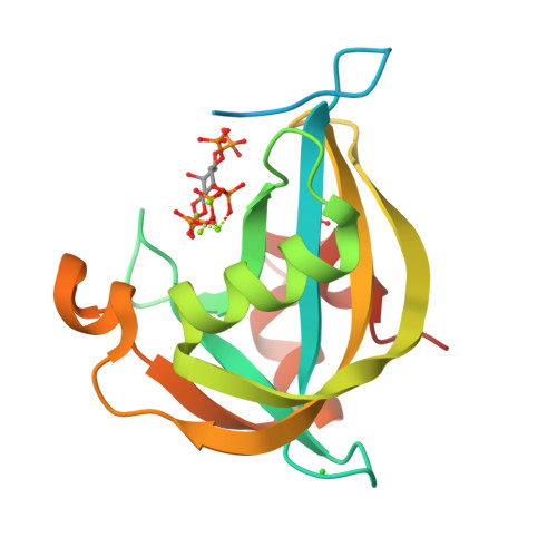
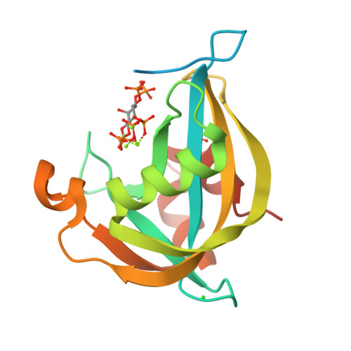
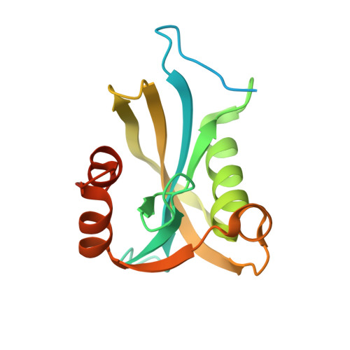
Diphosphoinositol polyphosphate phosphohydrolase 1 (DIPP1/NUDT3) in complex with 5-diphosphoinositol 1,3,4,6-tetrakisphosphate (5-PP-IP4), Mg, and Fluoride ion


Experimental Data Snapshot

  • Method: X-RAY DIFFRACTION
  • Resolution: 1.70 Å
  • R-Value Free: 0.216 
  • R-Value Work: 0.149 
  • R-Value Observed: 0.152 

wwPDB Validation   3D Report Full Report


Ligand Structure Quality Assessment 

Created with Raphaël 2.3.0Worse 01 BetterLigand structure goodness of fit to experimental dataBest fitted U6JClick on this verticalbar to view detailsBest fitted SO4Click on this verticalbar to view details

This is version 1.1 of the entry. See complete history


Literature

New structural insights reveal an expanded reaction cycle for inositol pyrophosphate hydrolysis by human DIPP1.

Zong, G.Jork, N.Hostachy, S.Fiedler, D.Jessen, H.J.Shears, S.B.Wang, H.

(2021) FASEB J 35: e21275-e21275

  • DOI: https://doi.org/10.1096/fj.202001489R
  • Primary Citation of Related Structures:  
    6WO7, 6WO8, 6WO9, 6WOA, 6WOB, 6WOC, 6WOD, 6WOE, 6WOF, 6WOG, 6WOH, 6WOI

  • PubMed Abstract: 

    Nudix hydrolases attract considerable attention for their wide range of specialized activities in all domains of life. One particular group of Nudix phosphohydrolases (DIPPs), through their metabolism of diphosphoinositol polyphosphates (PP-InsPs), regulates the actions of these polyphosphates upon bioenergetic homeostasis. In the current study, we describe, at an atomic level, hitherto unknown properties of human DIPP1.We provide X-ray analysis of the catalytic core of DIPP1 in crystals complexed with either natural PP-InsPs, alternative PP-InsP stereoisomers, or non-hydrolysable methylene bisphosphonate analogs ("PCP-InsPs"). The conclusions that we draw from these data are interrogated by studying the impact upon catalytic activity upon mutagenesis of certain key residues. We present a picture of a V-shaped catalytic furrow with overhanging ridges constructed from flexible positively charged side chains; within this cavity, the labile phosphoanhydride bond is appropriately positioned at the catalytic site by an extensive series of interlocking polar contacts which we analogize as "suspension cables." We demonstrate functionality for a triglycine peptide within a β-strand which represents a non-canonical addition to the standard Nudix catalytic core structure. We describe pre-reaction enzyme/substrate states which we posit to reflect a role for electrostatic steering in substrate capture. Finally, through time-resolved analysis, we uncover a chronological sequence of DIPP1/product post-reaction states, one of which may rationalize a role for InsP 6 as an inhibitor of catalytic activity.


  • Organizational Affiliation

    Signal Transduction Laboratory, National Institute of Environmental Health Sciences, National Institutes of Health, Research Triangle Park, NC, USA.


Macromolecules
Find similar proteins by:  (by identity cutoff)  |  3D Structure
Entity ID: 1
MoleculeChains Sequence LengthOrganismDetailsImage
Diphosphoinositol polyphosphate phosphohydrolase 1169Homo sapiensMutation(s): 0 
Gene Names: NUDT3DIPPDIPP1
EC: 3.6.1.52 (PDB Primary Data), 3.6.1 (PDB Primary Data), 3.6.1.62 (UniProt), 3.6.1.61 (UniProt), 3.6.1.59 (UniProt), 3.6.1.10 (UniProt)
UniProt & NIH Common Fund Data Resources
Find proteins for O95989 (Homo sapiens)
Explore O95989 
Go to UniProtKB:  O95989
PHAROS:  O95989
GTEx:  ENSG00000272325 
Entity Groups  
Sequence Clusters30% Identity50% Identity70% Identity90% Identity95% Identity100% Identity
UniProt GroupO95989
Sequence Annotations
Expand
  • Reference Sequence
Small Molecules
Ligands 5 Unique
IDChains Name / Formula / InChI Key2D Diagram3D Interactions
U6J (Subject of Investigation/LOI)
Query on U6J

Download Ideal Coordinates CCD File 
B [auth A](1r,2R,3S,4r,5R,6S)-4-hydroxy-2,3,5,6-tetrakis(phosphonooxy)cyclohexyl trihydrogen diphosphate
C6 H18 O24 P6
FFZGWHDHUIRNPY-KXXVROSKSA-N
SO4 (Subject of Investigation/LOI)
Query on SO4

Download Ideal Coordinates CCD File 
G [auth A]SULFATE ION
O4 S
QAOWNCQODCNURD-UHFFFAOYSA-L
CL (Subject of Investigation/LOI)
Query on CL

Download Ideal Coordinates CCD File 
F [auth A]CHLORIDE ION
Cl
VEXZGXHMUGYJMC-UHFFFAOYSA-M
MG (Subject of Investigation/LOI)
Query on MG

Download Ideal Coordinates CCD File 
C [auth A],
D [auth A],
E [auth A]
MAGNESIUM ION
Mg
JLVVSXFLKOJNIY-UHFFFAOYSA-N
F (Subject of Investigation/LOI)
Query on F

Download Ideal Coordinates CCD File 
H [auth A]FLUORIDE ION
F
KRHYYFGTRYWZRS-UHFFFAOYSA-M
Experimental Data & Validation

Experimental Data

  • Method: X-RAY DIFFRACTION
  • Resolution: 1.70 Å
  • R-Value Free: 0.216 
  • R-Value Work: 0.149 
  • R-Value Observed: 0.152 
  • Space Group: P 21 21 21
Unit Cell:
Length ( Å )Angle ( ˚ )
a = 46.28α = 90
b = 59.48β = 90
c = 62.26γ = 90
Software Package:
Software NamePurpose
REFMACrefinement
SCALEPACKdata scaling
PDB_EXTRACTdata extraction
DENZOdata reduction
PHASERphasing

Structure Validation

View Full Validation Report


Ligand Structure Quality Assessment 

Created with Raphaël 2.3.0Worse 01 BetterLigand structure goodness of fit to experimental dataBest fitted U6JClick on this verticalbar to view detailsBest fitted SO4Click on this verticalbar to view details

Entry History & Funding Information

Deposition Data


Funding OrganizationLocationGrant Number
National Institutes of Health/National Institute of Environmental Health Sciences (NIH/NIEHS)United States1ZIAES080046-31

Revision History  (Full details and data files)

  • Version 1.0: 2021-03-03
    Type: Initial release
  • Version 1.1: 2024-03-06
    Changes: Data collection, Database references