6XES

Tubulin-RB3_SLD in complex with compound 40a


Experimental Data Snapshot

  • Method: X-RAY DIFFRACTION
  • Resolution: 2.32 Å
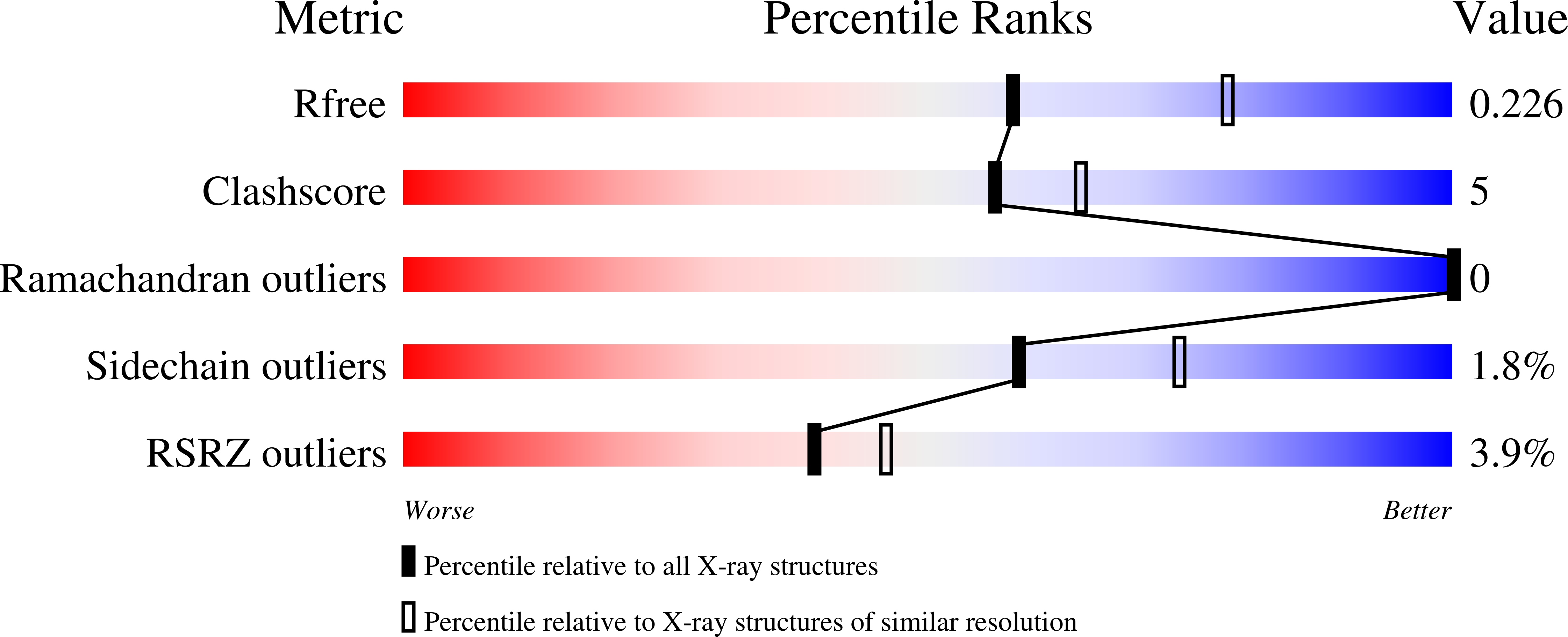
  • R-Value Free: 0.226 
  • R-Value Work: 0.171 
  • R-Value Observed: 0.173 

Starting Model: experimental
View more details

wwPDB Validation   3D Report Full Report


Ligand Structure Quality Assessment 


This is version 1.2 of the entry. See complete history


Literature

Design, Synthesis, and Biological Evaluation of Stable Colchicine-Binding Site Tubulin Inhibitors 6-Aryl-2-benzoyl-pyridines as Potential Anticancer Agents.

Chen, H.Deng, S.Albadari, N.Yun, M.K.Zhang, S.Li, Y.Ma, D.Parke, D.N.Yang, L.Seagroves, T.N.White, S.W.Miller, D.D.Li, W.

(2021) J Med Chem 64: 12049-12074

  • DOI: https://doi.org/10.1021/acs.jmedchem.1c00715
  • Primary Citation of Related Structures:  
    6XER, 6XES, 6XET

  • PubMed Abstract: 

    We previously reported a potent tubulin inhibitor CH-2-77. In this study, we optimized the structure of CH-2-77 by blocking metabolically labile sites and synthesized a series of CH-2-77 analogues. Two compounds, 40a and 60c , preserved the potency while improving the metabolic stability over CH-2-77 by 3- to 4-fold (46.8 and 29.4 vs 10.8 min in human microsomes). We determined the high-resolution X-ray crystal structures of 40a (resolution 2.3 Å) and 60c (resolution 2.6 Å) in complex with tubulin and confirmed their direct binding at the colchicine-binding site. In vitro , 60c maintained its mode of action by inhibiting tubulin polymerization and was effective against P-glycoprotein-mediated multiple drug resistance and taxol resistance. In vivo , 60c exhibited a strong inhibitory effect on tumor growth and metastasis in a taxol-resistant A375/TxR xenograft model without obvious toxicity. Collectively, this work showed that 60c is a promising lead compound for further development as a potential anticancer agent.


  • Organizational Affiliation

    Department of Pharmaceutical Sciences, College of Pharmacy, University of Tennessee Health Science Center, Memphis, Tennessee 38163, United States.


Macromolecules
Find similar proteins by:  (by identity cutoff)  |  3D Structure
Entity ID: 1
MoleculeChains Sequence LengthOrganismDetailsImage
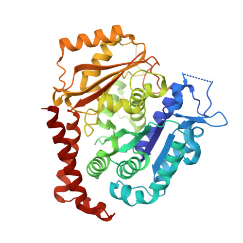
Tubulin alpha-1B chain
A, C
438Sus scrofaMutation(s): 0 
EC: 3.6.5
UniProt
Find proteins for Q2XVP4 (Sus scrofa)
Explore Q2XVP4 
Go to UniProtKB:  Q2XVP4
Entity Groups  
Sequence Clusters30% Identity50% Identity70% Identity90% Identity95% Identity100% Identity
UniProt GroupQ2XVP4
Sequence Annotations
Expand
  • Reference Sequence
Find similar proteins by:  (by identity cutoff)  |  3D Structure
Entity ID: 2
MoleculeChains Sequence LengthOrganismDetailsImage
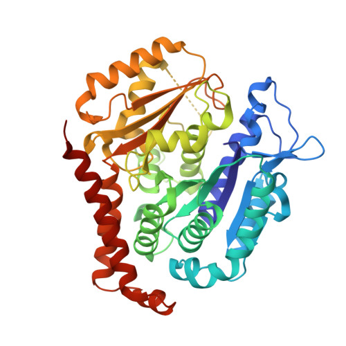
Tubulin beta chain
B, D
433Sus scrofaMutation(s): 0 
UniProt
Find proteins for P02554 (Sus scrofa)
Explore P02554 
Go to UniProtKB:  P02554
Entity Groups  
Sequence Clusters30% Identity50% Identity70% Identity90% Identity95% Identity100% Identity
UniProt GroupP02554
Sequence Annotations
Expand
  • Reference Sequence
Find similar proteins by:  (by identity cutoff)  |  3D Structure
Entity ID: 3
MoleculeChains Sequence LengthOrganismDetailsImage
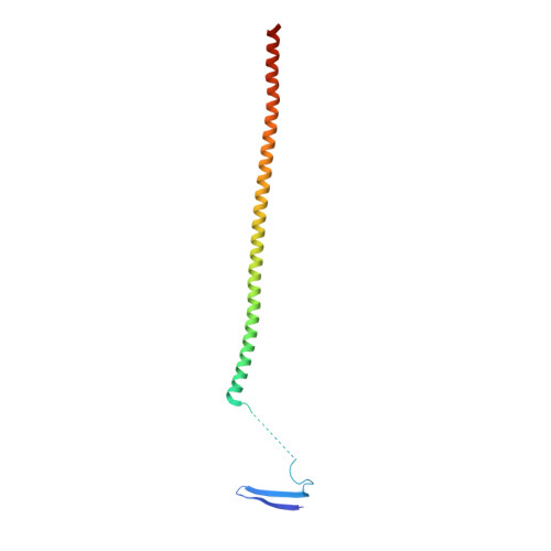
Stathmin-4143Rattus norvegicusMutation(s): 2 
Gene Names: Stmn4
UniProt
Find proteins for P63043 (Rattus norvegicus)
Explore P63043 
Go to UniProtKB:  P63043
Entity Groups  
Sequence Clusters30% Identity50% Identity70% Identity90% Identity95% Identity100% Identity
UniProt GroupP63043
Sequence Annotations
Expand
  • Reference Sequence
Small Molecules
Ligands 6 Unique
IDChains Name / Formula / InChI Key2D Diagram3D Interactions
GTP
Query on GTP

Download Ideal Coordinates CCD File 
F [auth A],
K [auth C],
M [auth D]
GUANOSINE-5'-TRIPHOSPHATE
C10 H16 N5 O14 P3
XKMLYUALXHKNFT-UUOKFMHZSA-N
GDP
Query on GDP

Download Ideal Coordinates CCD File 
H [auth B]GUANOSINE-5'-DIPHOSPHATE
C10 H15 N5 O11 P2
QGWNDRXFNXRZMB-UUOKFMHZSA-N
TU3 (Subject of Investigation/LOI)
Query on TU3

Download Ideal Coordinates CCD File 
J [auth B],
O [auth D]
[6-(3-hydroxy-4-methylphenyl)pyrazin-2-yl](3,4,5-trimethoxyphenyl)methanone
C21 H20 N2 O5
KWJXOYGITCRCRW-UHFFFAOYSA-N
SO4
Query on SO4

Download Ideal Coordinates CCD File 
G [auth A],
I [auth B],
N [auth D]
SULFATE ION
O4 S
QAOWNCQODCNURD-UHFFFAOYSA-L
DMS
Query on DMS

Download Ideal Coordinates CCD File 
P [auth D]DIMETHYL SULFOXIDE
C2 H6 O S
IAZDPXIOMUYVGZ-UHFFFAOYSA-N
MG
Query on MG

Download Ideal Coordinates CCD File 
L [auth C]MAGNESIUM ION
Mg
JLVVSXFLKOJNIY-UHFFFAOYSA-N
Experimental Data & Validation

Experimental Data

  • Method: X-RAY DIFFRACTION
  • Resolution: 2.32 Å
  • R-Value Free: 0.226 
  • R-Value Work: 0.171 
  • R-Value Observed: 0.173 
  • Space Group: P 21 21 21
Unit Cell:
Length ( Å )Angle ( ˚ )
a = 66.178α = 90
b = 126.966β = 90
c = 250.155γ = 90
Software Package:
Software NamePurpose
PHENIXrefinement
HKL-2000data reduction
HKL-2000data scaling
PHASERphasing

Structure Validation

View Full Validation Report


Ligand Structure Quality Assessment 


Entry History & Funding Information

Deposition Data


Funding OrganizationLocationGrant Number
National Institutes of Health/National Cancer Institute (NIH/NCI)United StatesR01CA148706

Revision History  (Full details and data files)

  • Version 1.0: 2021-08-25
    Type: Initial release
  • Version 1.1: 2021-09-08
    Changes: Database references
  • Version 1.2: 2023-10-18
    Changes: Data collection, Refinement description