7BZM

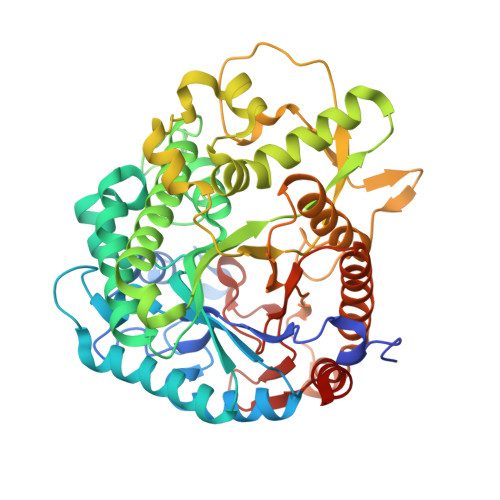
Crystal structure of rice Os3BGlu7 with glucoimidazole


Experimental Data Snapshot

  • Method: X-RAY DIFFRACTION
  • Resolution: 2.30 Å
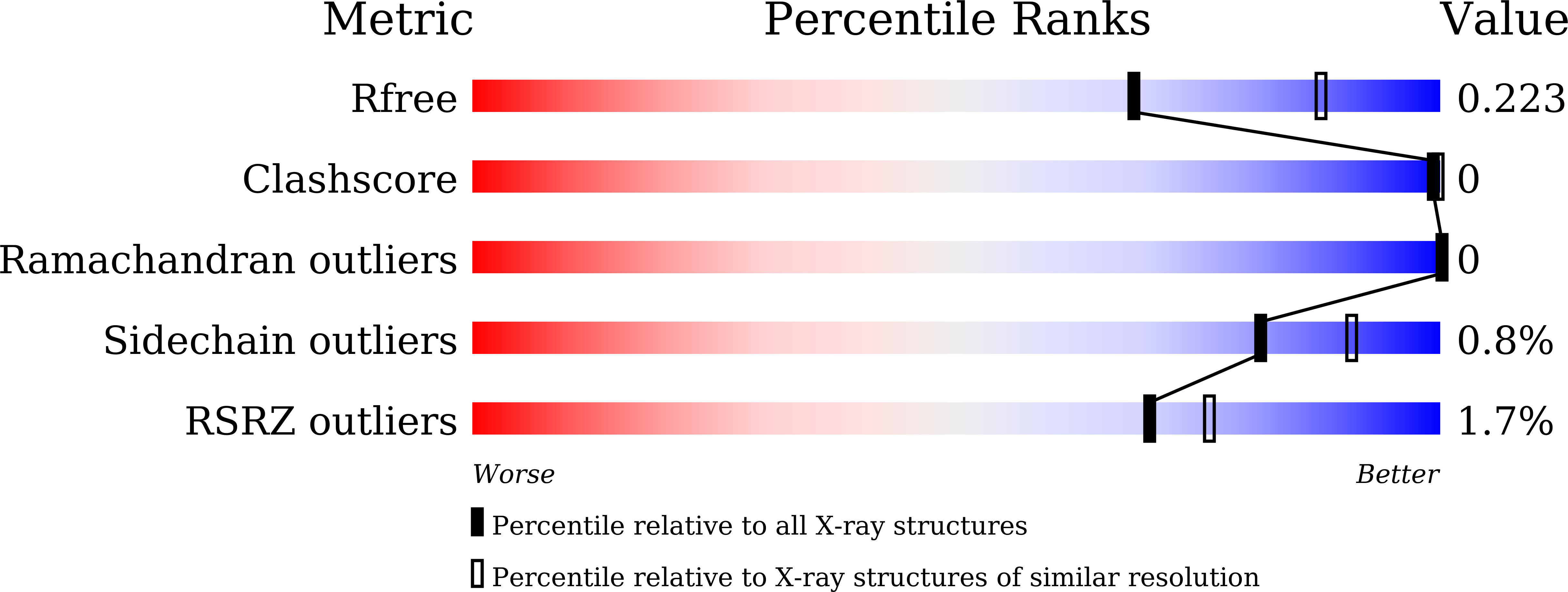
  • R-Value Free: 0.217 
  • R-Value Work: 0.177 
  • R-Value Observed: 0.179 

Starting Model: experimental
View more details

wwPDB Validation   3D Report Full Report


Ligand Structure Quality Assessment 


This is version 1.2 of the entry. See complete history


Literature

Structural Basis of Specific Glucoimidazole and Mannoimidazole Binding by Os3BGlu7.

Nutho, B.Pengthaisong, S.Tankrathok, A.Lee, V.S.Ketudat Cairns, J.R.Rungrotmongkol, T.Hannongbua, S.

(2020) Biomolecules 10

  • DOI: https://doi.org/10.3390/biom10060907
  • Primary Citation of Related Structures:  
    7BZM

  • PubMed Abstract: 

    β-Glucosidases and β-mannosidases hydrolyze substrates that differ only in the epimer of the nonreducing terminal sugar moiety, but most such enzymes show a strong preference for one activity or the other. Rice Os3BGlu7 and Os7BGlu26 β-glycosidases show a less strong preference, but Os3BGlu7 and Os7BGlu26 prefer glucosides and mannosides, respectively. Previous studies of crystal structures with glucoimidazole (GIm) and mannoimidazole (MIm) complexes and metadynamic simulations suggested that Os7BGlu26 hydrolyzes mannosides via the B 2,5 transition state (TS) conformation preferred for mannosides and glucosides via their preferred 4 H 3 / 4 E TS conformation. However, MIm is weakly bound by both enzymes. In the present study, we found that MIm was not bound in the active site of crystallized Os3BGlu7, but GIm was tightly bound in the -1 subsite in a 4 H 3 / 4 E conformation via hydrogen bonds with the surrounding residues. One-microsecond molecular dynamics simulations showed that GIm was stably bound in the Os3BGlu7 active site and the glycone-binding site with little distortion. In contrast, MIm initialized in the B 2,5 conformation rapidly relaxed to a E 3 / 4 H 3 conformation and moved out into a position in the entrance of the active site, where it bound more stably despite making fewer interactions. The lack of MIm binding in the glycone site in protein crystals and simulations implies that the energy required to distort MIm to the B 2,5 conformation for optimal active site residue interactions is sufficient to offset the energy of those interactions in Os3BGlu7. This balance between distortion and binding energy may also provide a rationale for glucosidase versus mannosidase specificity in plant β-glycosidases.


  • Organizational Affiliation

    Center of Excellence in Computational Chemistry (CECC), Department of Chemistry, Faculty of Science, Chulalongkorn University, Bangkok 10330, Thailand.


Macromolecules
Find similar proteins by:  (by identity cutoff)  |  3D Structure
Entity ID: 1
MoleculeChains Sequence LengthOrganismDetailsImage
Beta-glucosidase 7
A, B
481Oryza sativa Japonica GroupMutation(s): 0 
Gene Names: BGLU7BGLU1Os03g0703000LOC_Os03g49600OSJNBa0004L11.16
EC: 3.2.1.21
UniProt
Find proteins for Q75I93 (Oryza sativa subsp. japonica)
Explore Q75I93 
Go to UniProtKB:  Q75I93
Entity Groups  
Sequence Clusters30% Identity50% Identity70% Identity90% Identity95% Identity100% Identity
UniProt GroupQ75I93
Sequence Annotations
Expand
  • Reference Sequence
Small Molecules
Ligands 4 Unique
IDChains Name / Formula / InChI Key2D Diagram3D Interactions
GIM (Subject of Investigation/LOI)
Query on GIM

Download Ideal Coordinates CCD File 
C [auth A],
G [auth B]
GLUCOIMIDAZOLE
C8 H13 N2 O4
RZRDQZQPTISYKY-MVIOUDGNSA-O
MES (Subject of Investigation/LOI)
Query on MES

Download Ideal Coordinates CCD File 
D [auth A],
H [auth B]
2-(N-MORPHOLINO)-ETHANESULFONIC ACID
C6 H13 N O4 S
SXGZJKUKBWWHRA-UHFFFAOYSA-N
SO4 (Subject of Investigation/LOI)
Query on SO4

Download Ideal Coordinates CCD File 
F [auth A],
I [auth B]
SULFATE ION
O4 S
QAOWNCQODCNURD-UHFFFAOYSA-L
ZN (Subject of Investigation/LOI)
Query on ZN

Download Ideal Coordinates CCD File 
E [auth A]ZINC ION
Zn
PTFCDOFLOPIGGS-UHFFFAOYSA-N
Experimental Data & Validation

Experimental Data

  • Method: X-RAY DIFFRACTION
  • Resolution: 2.30 Å
  • R-Value Free: 0.217 
  • R-Value Work: 0.177 
  • R-Value Observed: 0.179 
  • Space Group: P 21 21 21
Unit Cell:
Length ( Å )Angle ( ˚ )
a = 80.402α = 90
b = 101.557β = 90
c = 127.458γ = 90
Software Package:
Software NamePurpose
REFMACrefinement
HKL-2000data reduction
HKL-2000data scaling
MOLREPphasing
PDB_EXTRACTdata extraction
Cootmodel building

Structure Validation

View Full Validation Report


Ligand Structure Quality Assessment 


Entry History & Funding Information

Deposition Data


Funding OrganizationLocationGrant Number
Other governmentThailandBRG5980015 Thailand Research Fund and Suranaree University of Technology
Other governmentThailandFtR.15/2560 Suranaree University of Technology and Office of the Higher Education Commission under NRU Project of Thailand

Revision History  (Full details and data files)

  • Version 1.0: 2020-07-08
    Type: Initial release
  • Version 1.1: 2023-11-29
    Changes: Data collection, Database references, Derived calculations, Refinement description
  • Version 1.2: 2024-10-16
    Changes: Structure summary