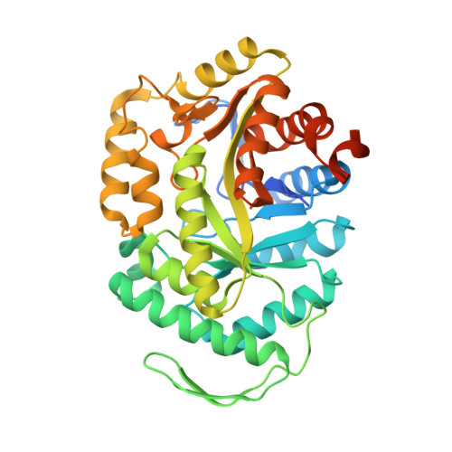
7K14

Ternary soak structure of alkanesulfonate monooxygenase MsuD from Pseudomonas fluorescens with FMN and methanesulfonate


Experimental Data Snapshot

  • Method: X-RAY DIFFRACTION
  • Resolution: 2.75 Å
  • R-Value Free: 0.214 
  • R-Value Work: 0.172 
  • R-Value Observed: 0.174 

Starting Model: experimental
View more details

wwPDB Validation   3D Report Full Report


Ligand Structure Quality Assessment 


This is version 1.3 of the entry. See complete history


Literature

Structures of the alkanesulfonate monooxygenase MsuD provide insight into C-S bond cleavage, substrate scope, and an unexpected role for the tetramer.

Liew, J.J.M.El Saudi, I.M.Nguyen, S.V.Wicht, D.K.Dowling, D.P.

(2021) J Biol Chem 297: 100823-100823

  • DOI: https://doi.org/10.1016/j.jbc.2021.100823
  • Primary Citation of Related Structures:  
    7JV3, 7JW9, 7JYB, 7K14, 7K64

  • PubMed Abstract: 

    Bacterial two-component flavin-dependent monooxygenases cleave the stable C-S bond of environmental and anthropogenic organosulfur compounds. The monooxygenase MsuD converts methanesulfonate (MS - ) to sulfite, completing the sulfur assimilation process during sulfate starvation, but the mechanism of this conversion remains unclear. To explore the mechanism of C-S bond cleavage, we report a series of crystal structures of MsuD from Pseudomonas fluorescens in different liganded states. This report provides the first crystal structures of an alkanesulfonate monooxygenase with a bound flavin and alkanesulfonate, elucidating the roles of the active site lid, the protein C terminus, and an active site loop in flavin and/or alkanesulfonate binding. These structures position MS - closest to the flavin N5 position, consistent with an N5-(hydro)peroxyflavin mechanism rather than a classical C4a-(hydro)peroxyflavin mechanism. A fully enclosed active site is observed in the ternary complex, mediated by interchain interaction of the C terminus at the tetramer interface. These structures identify an unexpected function of the protein C terminus in this protein family in stabilizing tetramer formation and the alkanesulfonate-binding site. Spurred by interest from the crystal structures, we conducted biochemical assays and molecular docking that redefine MsuD as a small- to medium-chain alkanesulfonate monooxygenase. Functional mutations verify the sulfonate-binding site and reveal the critical importance of the protein C terminus for monooxygenase function. These findings reveal a deeper understanding of MsuD's functionality at the molecular level and consequently how it operates within its role as part of the sulfur assimilation pathway.


  • Organizational Affiliation

    Department of Chemistry, University of Massachusetts Boston, Boston, Massachusetts, USA.


Macromolecules
Find similar proteins by:  (by identity cutoff)  |  3D Structure
Entity ID: 1
MoleculeChains Sequence LengthOrganismDetailsImage
Alkanesulfonate monooxygenase
A, B, C, D, E
A, B, C, D, E, F, G, H
404Pseudomonas fluorescens Pf0-1Mutation(s): 0 
Gene Names: msuDssuDPfl01_3916
EC: 1.14.14.5
UniProt
Find proteins for Q3K9A1 (Pseudomonas fluorescens (strain Pf0-1))
Explore Q3K9A1 
Go to UniProtKB:  Q3K9A1
Entity Groups  
Sequence Clusters30% Identity50% Identity70% Identity90% Identity95% Identity100% Identity
UniProt GroupQ3K9A1
Sequence Annotations
Expand
  • Reference Sequence
Small Molecules
Ligands 4 Unique
IDChains Name / Formula / InChI Key2D Diagram3D Interactions
FMN (Subject of Investigation/LOI)
Query on FMN

Download Ideal Coordinates CCD File 
I [auth A]
K [auth B]
M [auth C]
O [auth D]
Q [auth E]
I [auth A],
K [auth B],
M [auth C],
O [auth D],
Q [auth E],
T [auth G],
V [auth H]
FLAVIN MONONUCLEOTIDE
C17 H21 N4 O9 P
FVTCRASFADXXNN-SCRDCRAPSA-N
03S
Query on 03S

Download Ideal Coordinates CCD File 
J [auth A],
L [auth B],
N [auth C],
P [auth D],
R [auth E]
methanesulfonic acid
C H4 O3 S
AFVFQIVMOAPDHO-UHFFFAOYSA-N
PO4
Query on PO4

Download Ideal Coordinates CCD File 
S [auth F]PHOSPHATE ION
O4 P
NBIIXXVUZAFLBC-UHFFFAOYSA-K
CL
Query on CL

Download Ideal Coordinates CCD File 
U [auth G],
W [auth H]
CHLORIDE ION
Cl
VEXZGXHMUGYJMC-UHFFFAOYSA-M
Experimental Data & Validation

Experimental Data

  • Method: X-RAY DIFFRACTION
  • Resolution: 2.75 Å
  • R-Value Free: 0.214 
  • R-Value Work: 0.172 
  • R-Value Observed: 0.174 
  • Space Group: P 1 21 1
Unit Cell:
Length ( Å )Angle ( ˚ )
a = 94α = 90
b = 211.93β = 119.049
c = 94.35γ = 90
Software Package:
Software NamePurpose
PHENIXrefinement
XDSdata reduction
XSCALEdata scaling
PHASERphasing

Structure Validation

View Full Validation Report


Ligand Structure Quality Assessment 


Entry History & Funding Information

Deposition Data


Funding OrganizationLocationGrant Number
National Science Foundation (NSF, United States)United States1807480

Revision History  (Full details and data files)

  • Version 1.0: 2021-05-26
    Type: Initial release
  • Version 1.1: 2021-06-09
    Changes: Database references
  • Version 1.2: 2021-07-07
    Changes: Database references
  • Version 1.3: 2023-10-18
    Changes: Data collection, Database references, Refinement description