7MOG

Crystal Structure of Arabidopsis thaliana Plant and Fungi Atypical Dual Specificity Phosphatase 1(AtPFA-DSP1 ) Cys150Ser in complex with 5-PCF2 Am-InsP5, an analogue of 5-InsP7

  • Classification: HYDROLASE
  • Organism(s): Arabidopsis thaliana
  • Expression System: Escherichia coli
  • Mutation(s): Yes 

  • Deposited: 2021-05-01 Released: 2022-03-02 
  • Deposition Author(s): Wang, H., Shears, S.B.
  • Funding Organization(s): National Institutes of Health/National Institute of Environmental Health Sciences (NIH/NIEHS)

Experimental Data Snapshot

  • Method: X-RAY DIFFRACTION
  • Resolution: 1.80 Å
  • R-Value Free: 
    0.147 (Depositor), 0.150 (DCC) 
  • R-Value Work: 
    0.119 (Depositor), 0.120 (DCC) 
  • R-Value Observed: 
    0.120 (Depositor) 

wwPDB Validation   3D Report Full Report


Ligand Structure Quality Assessment 

Created with Raphaël 2.3.0Worse 01 BetterLigand structure goodness of fit to experimental dataBest fitted KDJClick on this verticalbar to view details

This is version 1.2 of the entry. See complete history


Literature

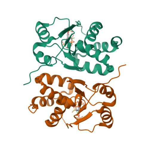
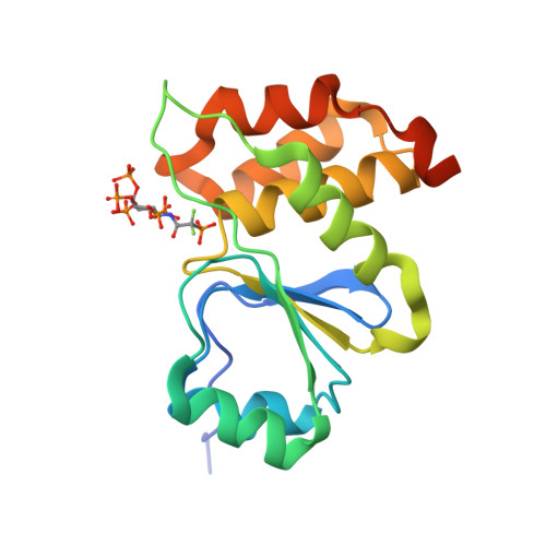
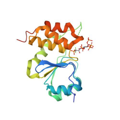
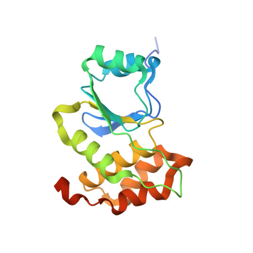
A structural expose of noncanonical molecular reactivity within the protein tyrosine phosphatase WPD loop.

Wang, H.Perera, L.Jork, N.Zong, G.Riley, A.M.Potter, B.V.L.Jessen, H.J.Shears, S.B.

(2022) Nat Commun 13: 2231-2231

  • DOI: https://doi.org/10.1038/s41467-022-29673-y
  • Primary Citation of Related Structures:  
    7MOD, 7MOE, 7MOF, 7MOG, 7MOH, 7MOI, 7MOJ, 7MOK, 7MOL, 7MOM

  • PubMed Abstract: 

    Structural snapshots of protein/ligand complexes are a prerequisite for gaining atomic level insight into enzymatic reaction mechanisms. An important group of enzymes has been deprived of this analytical privilege: members of the protein tyrosine phosphatase (PTP) superfamily with catalytic WPD-loops lacking the indispensable general-acid/base within a tryptophan-proline-aspartate/glutamate context. Here, we provide the ligand/enzyme crystal complexes for one such PTP outlier: Arabidopsis thaliana Plant and Fungi Atypical Dual Specificity Phosphatase 1 (AtPFA-DSP1), herein unveiled as a regioselective and efficient phosphatase towards inositol pyrophosphate (PP-InsP) signaling molecules. Although the WPD loop is missing its canonical tripeptide motif, this structural element contributes to catalysis by assisting PP-InsP delivery into the catalytic pocket, for a choreographed exchange with phosphate reaction product. Subsequently, an intramolecular proton donation by PP-InsP substrate is posited to substitute functionally for the absent aspartate/glutamate general-acid. Overall, we expand mechanistic insight into adaptability of the conserved PTP structural elements.


  • Organizational Affiliation

    Signal Transduction Laboratory, National Institute of Environmental Health Sciences, National Institutes of Health, Research Triangle Park, NC, 27709, USA. huanchen.wang@nih.gov.


Macromolecules
Find similar proteins by:  (by identity cutoff)  |  3D Structure
Entity ID: 1
MoleculeChains Sequence LengthOrganismDetailsImage
Tyrosine-protein phosphatase DSP1
A, B
171Arabidopsis thalianaMutation(s): 1 
Gene Names: DSP1PTP135At1g05000T7A14.14
EC: 3.1.3.48 (PDB Primary Data), 3.6.1.52 (UniProt)
UniProt
Find proteins for Q9ZVN4 (Arabidopsis thaliana)
Explore Q9ZVN4 
Go to UniProtKB:  Q9ZVN4
Entity Groups  
Sequence Clusters30% Identity50% Identity70% Identity90% Identity95% Identity100% Identity
UniProt GroupQ9ZVN4
Sequence Annotations
Expand
  • Reference Sequence
Small Molecules
Ligands 1 Unique
IDChains Name / Formula / InChI Key2D Diagram3D Interactions
KDJ (Subject of Investigation/LOI)
Query on KDJ

Download Ideal Coordinates CCD File 
C [auth A],
D [auth B]
(1,1-difluoro-2-oxo-2-{[(1s,2R,3S,4s,5R,6S)-2,3,4,5,6-pentakis(phosphonooxy)cyclohexyl]amino}ethyl)phosphonic acid
C8 H19 F2 N O24 P6
XBHZOGSBYRIXJA-QWBQGLJISA-N
Experimental Data & Validation

Experimental Data

  • Method: X-RAY DIFFRACTION
  • Resolution: 1.80 Å
  • R-Value Free:  0.147 (Depositor), 0.150 (DCC) 
  • R-Value Work:  0.119 (Depositor), 0.120 (DCC) 
  • R-Value Observed: 0.120 (Depositor) 
Space Group: P 21 3
Unit Cell:
Length ( Å )Angle ( ˚ )
a = 124.654α = 90
b = 124.654β = 90
c = 124.654γ = 90
Software Package:
Software NamePurpose
REFMACrefinement
HKL-2000data reduction
HKL-2000data scaling
PDB_EXTRACTdata extraction
REFMACphasing

Structure Validation

View Full Validation Report


Ligand Structure Quality Assessment 

Created with Raphaël 2.3.0Worse 01 BetterLigand structure goodness of fit to experimental dataBest fitted KDJClick on this verticalbar to view details

Entry History & Funding Information

Deposition Data


Funding OrganizationLocationGrant Number
National Institutes of Health/National Institute of Environmental Health Sciences (NIH/NIEHS)United States1ZIAES080046-29

Revision History  (Full details and data files)

  • Version 1.0: 2022-03-02
    Type: Initial release
  • Version 1.1: 2022-05-11
    Changes: Database references
  • Version 1.2: 2024-05-22
    Changes: Data collection