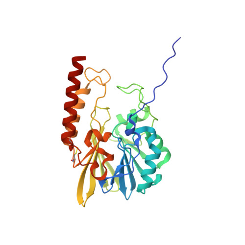
7O0O

Crystal structure of the B3 metallo-beta-lactamase L1 with hydrolysed ertapenem


Experimental Data Snapshot

  • Method: X-RAY DIFFRACTION
  • Resolution: 1.45 Å
  • R-Value Free: 0.169 
  • R-Value Work: 0.154 
  • R-Value Observed: 0.155 

Starting Model: experimental
View more details

wwPDB Validation   3D Report Full Report


Ligand Structure Quality Assessment 


This is version 2.3 of the entry. See complete history


Literature

Crystallography and QM/MM Simulations Identify Preferential Binding of Hydrolyzed Carbapenem and Penem Antibiotics to the L1 Metallo-beta-Lactamase in the Imine Form.

Twidale, R.M.Hinchliffe, P.Spencer, J.Mulholland, A.J.

(2021) J Chem Inf Model 61: 5988-5999

  • DOI: https://doi.org/10.1021/acs.jcim.1c00663
  • Primary Citation of Related Structures:  
    7O0O

  • PubMed Abstract: 

    Widespread bacterial resistance to carbapenem antibiotics is an increasing global health concern. Resistance has emerged due to carbapenem-hydrolyzing enzymes, including metallo-β-lactamases (MβLs), but despite their prevalence and clinical importance, MβL mechanisms are still not fully understood. Carbapenem hydrolysis by MβLs can yield alternative product tautomers with the potential to access different binding modes. Here, we show that a combined approach employing crystallography and quantum mechanics/molecular mechanics (QM/MM) simulations allow tautomer assignment in MβL:hydrolyzed antibiotic complexes. Molecular simulations also examine (meta)stable species of alternative protonation and tautomeric states, providing mechanistic insights into β-lactam hydrolysis. We report the crystal structure of the hydrolyzed carbapenem ertapenem bound to the L1 MβL from Stenotrophomonas maltophilia and model alternative tautomeric and protonation states of both hydrolyzed ertapenem and faropenem (a related penem antibiotic), which display different binding modes with L1. We show how the structures of both complexed β-lactams are best described as the (2 S )-imine tautomer with the carboxylate formed after β-lactam ring cleavage deprotonated. Simulations show that enamine tautomer complexes are significantly less stable (e.g., showing partial loss of interactions with the L1 binuclear zinc center) and not consistent with experimental data. Strong interactions of Tyr32 and one zinc ion (Zn1) with ertapenem prevent a C6 group rotation, explaining the different binding modes of the two β-lactams. Our findings establish the relative stability of different hydrolyzed (carba)penem forms in the L1 active site and identify interactions important to stable complex formation, information that should assist inhibitor design for this important antibiotic resistance determinant.


  • Organizational Affiliation

    Centre for Computational Chemistry, School of Chemistry, University of Bristol, Cantock's Close, Bristol BS8 1TS, U.K.


Macromolecules
Find similar proteins by:  (by identity cutoff)  |  3D Structure
Entity ID: 1
MoleculeChains Sequence LengthOrganismDetailsImage
Metallo-beta-lactamase L1271Stenotrophomonas maltophiliaMutation(s): 0 
EC: 3.5.2.6
UniProt
Find proteins for P52700 (Stenotrophomonas maltophilia)
Explore P52700 
Go to UniProtKB:  P52700
Entity Groups  
Sequence Clusters30% Identity50% Identity70% Identity90% Identity95% Identity100% Identity
UniProt GroupP52700
Sequence Annotations
Expand
  • Reference Sequence
Small Molecules
Ligands 3 Unique
IDChains Name / Formula / InChI Key2D Diagram3D Interactions
9XS (Subject of Investigation/LOI)
Query on 9XS

Download Ideal Coordinates CCD File 
E [auth A](2~{S},3~{R},4~{S})-2-[(2~{S},3~{R})-1,3-bis(oxidanyl)-1-oxidanylidene-butan-2-yl]-4-[(3~{S},5~{S})-5-[(3-carboxyphenyl)carbamoyl]pyrrolidin-3-yl]sulfanyl-3-methyl-3,4-dihydro-2~{H}-pyrrole-5-carboxylic acid
C22 H27 N3 O8 S
MLCVCXYVRPDWBV-UIRAGYLNSA-N
SO4
Query on SO4

Download Ideal Coordinates CCD File 
F [auth A],
G [auth A]
SULFATE ION
O4 S
QAOWNCQODCNURD-UHFFFAOYSA-L
ZN
Query on ZN

Download Ideal Coordinates CCD File 
B [auth A],
C [auth A],
D [auth A]
ZINC ION
Zn
PTFCDOFLOPIGGS-UHFFFAOYSA-N
Experimental Data & Validation

Experimental Data

  • Method: X-RAY DIFFRACTION
  • Resolution: 1.45 Å
  • R-Value Free: 0.169 
  • R-Value Work: 0.154 
  • R-Value Observed: 0.155 
  • Space Group: P 64 2 2
Unit Cell:
Length ( Å )Angle ( ˚ )
a = 105.803α = 90
b = 105.803β = 90
c = 98.149γ = 120
Software Package:
Software NamePurpose
PHENIXrefinement
PDB_EXTRACTdata extraction
DIALSdata reduction
DIALSdata scaling
PHASERphasing

Structure Validation

View Full Validation Report


Ligand Structure Quality Assessment 


Entry History & Funding Information

Deposition Data


Funding OrganizationLocationGrant Number
National Institutes of Health/National Institute Of Allergy and Infectious Diseases (NIH/NIAID)United StatesR01AI100560

Revision History  (Full details and data files)

  • Version 1.0: 2021-10-13
    Type: Initial release
  • Version 2.0: 2021-12-01
    Changes: Advisory, Atomic model, Author supporting evidence, Data collection, Derived calculations, Non-polymer description, Structure summary
  • Version 2.1: 2022-02-16
    Changes: Database references, Derived calculations
  • Version 2.2: 2024-01-31
    Changes: Data collection, Refinement description
  • Version 2.3: 2024-10-23
    Changes: Structure summary