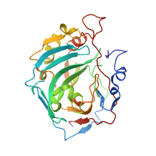
7Q0C

Mimic carbonic anhydrase IX in complex with Methyl 2-chloro-4-(cyclohexylsulfanyl)-5-sulfamoylbenzoate


Experimental Data Snapshot

  • Method: X-RAY DIFFRACTION
  • Resolution: 1.12 Å
  • R-Value Free: 0.194 
  • R-Value Work: 0.160 
  • R-Value Observed: 0.164 

Starting Model: experimental
View more details

wwPDB Validation   3D Report Full Report


Ligand Structure Quality Assessment 


This is version 1.2 of the entry. See complete history


Literature

Methyl 2-Halo-4-Substituted-5-Sulfamoyl-Benzoates as High Affinity and Selective Inhibitors of Carbonic Anhydrase IX.

Zaksauskas, A.Capkauskaite, E.Paketuryte-Latve, V.Smirnov, A.Leitans, J.Kazaks, A.Dvinskis, E.Stancaitis, L.Mickeviciute, A.Jachno, J.Jezepcikas, L.Linkuviene, V.Sakalauskas, A.Manakova, E.Grazulis, S.Matuliene, J.Tars, K.Matulis, D.

(2021) Int J Mol Sci 23

  • DOI: https://doi.org/10.3390/ijms23010130
  • Primary Citation of Related Structures:  
    7POM, 7PP9, 7PUU, 7PUV, 7PUW, 7Q0C, 7Q0D, 7Q0E

  • PubMed Abstract: 

    Among the twelve catalytically active carbonic anhydrase isozymes present in the human body, the CAIX is highly overexpressed in various solid tumors. The enzyme acidifies the tumor microenvironment enabling invasion and metastatic processes. Therefore, many attempts have been made to design chemical compounds that would exhibit high affinity and selective binding to CAIX over the remaining eleven catalytically active CA isozymes to limit undesired side effects. It has been postulated that such drugs may have anticancer properties and could be used in tumor treatment. Here we have designed a series of compounds, methyl 5-sulfamoyl-benzoates, which bear a primary sulfonamide group, a well-known marker of CA inhibitors, and determined their affinities for all twelve CA isozymes. Variations of substituents on the benzenesulfonamide ring led to compound 4b , which exhibited an extremely high observed binding affinity to CAIX; the K d was 0.12 nM. The intrinsic dissociation constant, where the binding-linked protonation reactions have been subtracted, reached 0.08 pM. The compound also exhibited more than 100-fold selectivity over the remaining CA isozymes. The X-ray crystallographic structure of compound 3b bound to CAIX showed the structural position, while several structures of compounds bound to other CA isozymes showed structural reasons for compound selectivity towards CAIX. Since this series of compounds possess physicochemical properties suitable for drugs, they may be developed for anticancer therapeutic purposes.


  • Organizational Affiliation

    Department of Biothermodynamics and Drug Design, Institute of Biotechnology, Life Sciences Center, Vilnius University, Saulėtekio al. 7, LT-10257 Vilnius, Lithuania.


Macromolecules
Find similar proteins by:  (by identity cutoff)  |  3D Structure
Entity ID: 1
MoleculeChains Sequence LengthOrganismDetailsImage
Carbonic anhydrase 2
A, B
260Homo sapiensMutation(s): 6 
Gene Names: CA2
EC: 4.2.1.1 (PDB Primary Data), 4.2.1.69 (UniProt)
UniProt & NIH Common Fund Data Resources
Find proteins for P00918 (Homo sapiens)
Explore P00918 
Go to UniProtKB:  P00918
PHAROS:  P00918
GTEx:  ENSG00000104267 
Entity Groups  
Sequence Clusters30% Identity50% Identity70% Identity90% Identity95% Identity100% Identity
UniProt GroupP00918
Sequence Annotations
Expand
  • Reference Sequence
Small Molecules
Ligands 4 Unique
IDChains Name / Formula / InChI Key2D Diagram3D Interactions
7VZ (Subject of Investigation/LOI)
Query on 7VZ

Download Ideal Coordinates CCD File 
D [auth A],
H [auth B]
methyl 2-chloranyl-4-cyclohexylsulfanyl-5-sulfamoyl-benzoate
C14 H18 Cl N O4 S2
QYEZJVLETJURBN-UHFFFAOYSA-N
DMS
Query on DMS

Download Ideal Coordinates CCD File 
F [auth A],
I [auth B]
DIMETHYL SULFOXIDE
C2 H6 O S
IAZDPXIOMUYVGZ-UHFFFAOYSA-N
ZN
Query on ZN

Download Ideal Coordinates CCD File 
C [auth A],
G [auth B]
ZINC ION
Zn
PTFCDOFLOPIGGS-UHFFFAOYSA-N
ACT
Query on ACT

Download Ideal Coordinates CCD File 
E [auth A]ACETATE ION
C2 H3 O2
QTBSBXVTEAMEQO-UHFFFAOYSA-M
Experimental Data & Validation

Experimental Data

  • Method: X-RAY DIFFRACTION
  • Resolution: 1.12 Å
  • R-Value Free: 0.194 
  • R-Value Work: 0.160 
  • R-Value Observed: 0.164 
  • Space Group: P 1
Unit Cell:
Length ( Å )Angle ( ˚ )
a = 42.06α = 90
b = 41.348β = 104.14
c = 72.029γ = 90
Software Package:
Software NamePurpose
REFMACrefinement
SCALAdata scaling
PDB_EXTRACTdata extraction
XDSdata reduction
MOLREPphasing
Cootmodel building

Structure Validation

View Full Validation Report


Ligand Structure Quality Assessment 


Entry History & Funding Information

Deposition Data


Funding OrganizationLocationGrant Number
Not funded--

Revision History  (Full details and data files)

  • Version 1.0: 2022-01-12
    Type: Initial release
  • Version 1.1: 2022-01-26
    Changes: Database references
  • Version 1.2: 2024-01-31
    Changes: Data collection, Refinement description