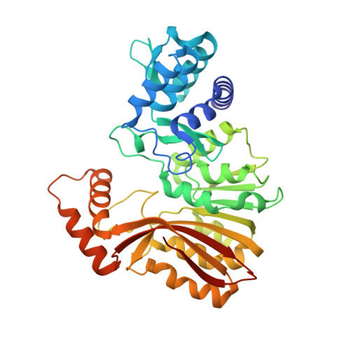
7RC2

Aeronamide N-methyltransferase, AerE


Experimental Data Snapshot

  • Method: X-RAY DIFFRACTION
  • Resolution: 1.51 Å
  • R-Value Free: 0.175 
  • R-Value Work: 0.161 
  • R-Value Observed: 0.162 

wwPDB Validation   3D Report Full Report


Ligand Structure Quality Assessment 


This is version 1.1 of the entry. See complete history


Literature

Structure and mechanism for iterative amide N -methylation in the biosynthesis of channel-forming peptide cytotoxins.

Cogan, D.P.Bhushan, A.Reyes, R.Zhu, L.Piel, J.Nair, S.K.

(2022) Proc Natl Acad Sci U S A 119: e2116578119-e2116578119

  • DOI: https://doi.org/10.1073/pnas.2116578119
  • Primary Citation of Related Structures:  
    7RC2, 7RC3, 7RC4, 7RC5, 7RC6

  • PubMed Abstract: 

    SignificanceThe channel-forming proteusins are bacterial helical peptides that allow permeation of positively charged ions to influence membrane potential and cellular physiology. We biochemically characterize the effect of two critical posttranslational modifications on the secondary structure of the peptide substrate. We determine how a methyl group can be added to the side chains of D-Asn residues in a peptide substrate and show how flanking residues influence selectivity. These studies should foster the development of small-molecule peptide ion channels as therapeutics.


  • Organizational Affiliation

    Department of Biochemistry, University of Illinois at Urbana-Champaign, Urbana, IL 61801.


Macromolecules
Find similar proteins by:  (by identity cutoff)  |  3D Structure
Entity ID: 1
MoleculeChains Sequence LengthOrganismDetailsImage
Methyltransferase family protein384Microvirgula aerodenitrificans DSM 15089Mutation(s): 0 
Gene Names: aerE
UniProt
Find proteins for A0A2S0PEL5 (Microvirgula aerodenitrificans)
Explore A0A2S0PEL5 
Go to UniProtKB:  A0A2S0PEL5
Entity Groups  
Sequence Clusters30% Identity50% Identity70% Identity90% Identity95% Identity100% Identity
UniProt GroupA0A2S0PEL5
Sequence Annotations
Expand
  • Reference Sequence
Experimental Data & Validation

Experimental Data

  • Method: X-RAY DIFFRACTION
  • Resolution: 1.51 Å
  • R-Value Free: 0.175 
  • R-Value Work: 0.161 
  • R-Value Observed: 0.162 
  • Space Group: P 32 2 1
Unit Cell:
Length ( Å )Angle ( ˚ )
a = 77.566α = 90
b = 77.566β = 90
c = 152.314γ = 120
Software Package:
Software NamePurpose
PHENIXrefinement
PDB_EXTRACTdata extraction
XDSdata reduction
Aimlessdata scaling
AutoSolphasing

Structure Validation

View Full Validation Report


Ligand Structure Quality Assessment 


Entry History 

Deposition Data

Revision History  (Full details and data files)

  • Version 1.0: 2022-03-30
    Type: Initial release
  • Version 1.1: 2024-05-22
    Changes: Data collection