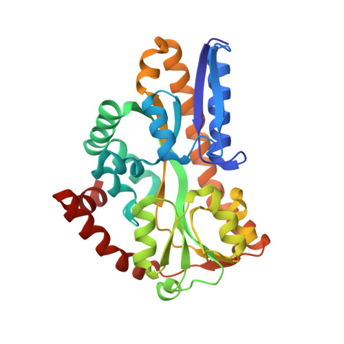
Structure and mechanism of a tripartite ATP-independent periplasmic TRAP transporter.
Davies, J.S., Currie, M.J., North, R.A., Scalise, M., Wright, J.D., Copping, J.M., Remus, D.M., Gulati, A., Morado, D.R., Jamieson, S.A., Newton-Vesty, M.C., Abeysekera, G.S., Ramaswamy, S., Friemann, R., Wakatsuki, S., Allison, J.R., Indiveri, C., Drew, D., Mace, P.D., Dobson, R.C.J.(2023) Nat Commun 14: 1120-1120
- PubMed: 36849793
- DOI: https://doi.org/10.1038/s41467-023-36590-1
- Primary Citation of Related Structures:
7QHA, 7T3E, 8B01 - PubMed Abstract:
In bacteria and archaea, tripartite ATP-independent periplasmic (TRAP) transporters uptake essential nutrients. TRAP transporters receive their substrates via a secreted soluble substrate-binding protein. How a sodium ion-driven secondary active transporter is strictly coupled to a substrate-binding protein is poorly understood. Here we report the cryo-EM structure of the sialic acid TRAP transporter SiaQM from Photobacterium profundum at 2.97 Å resolution. SiaM comprises a "transport" domain and a "scaffold" domain, with the transport domain consisting of helical hairpins as seen in the sodium ion-coupled elevator transporter VcINDY. The SiaQ protein forms intimate contacts with SiaM to extend the size of the scaffold domain, suggesting that TRAP transporters may operate as monomers, rather than the typically observed oligomers for elevator-type transporters. We identify the Na + and sialic acid binding sites in SiaM and demonstrate a strict dependence on the substrate-binding protein SiaP for uptake. We report the SiaP crystal structure that, together with docking studies, suggest the molecular basis for how sialic acid is delivered to the SiaQM transporter complex. We thus propose a model for substrate transport by TRAP proteins, which we describe herein as an 'elevator-with-an-operator' mechanism.
Organizational Affiliation:
Biomolecular Interaction Centre, Maurice Wilkins Centre for Biodiscovery, MacDiarmid Institute for Advanced Materials and Nanotechnology and School of Biological Sciences, University of Canterbury, PO Box 4800, Christchurch, 8140, New Zealand.


