8B05

TRYPTOPHAN SYNTHASE - Cryo-trapping by the spitrobot crystal plunger after 20 sec


Experimental Data Snapshot

  • Method: X-RAY DIFFRACTION
  • Resolution: 2.10 Å
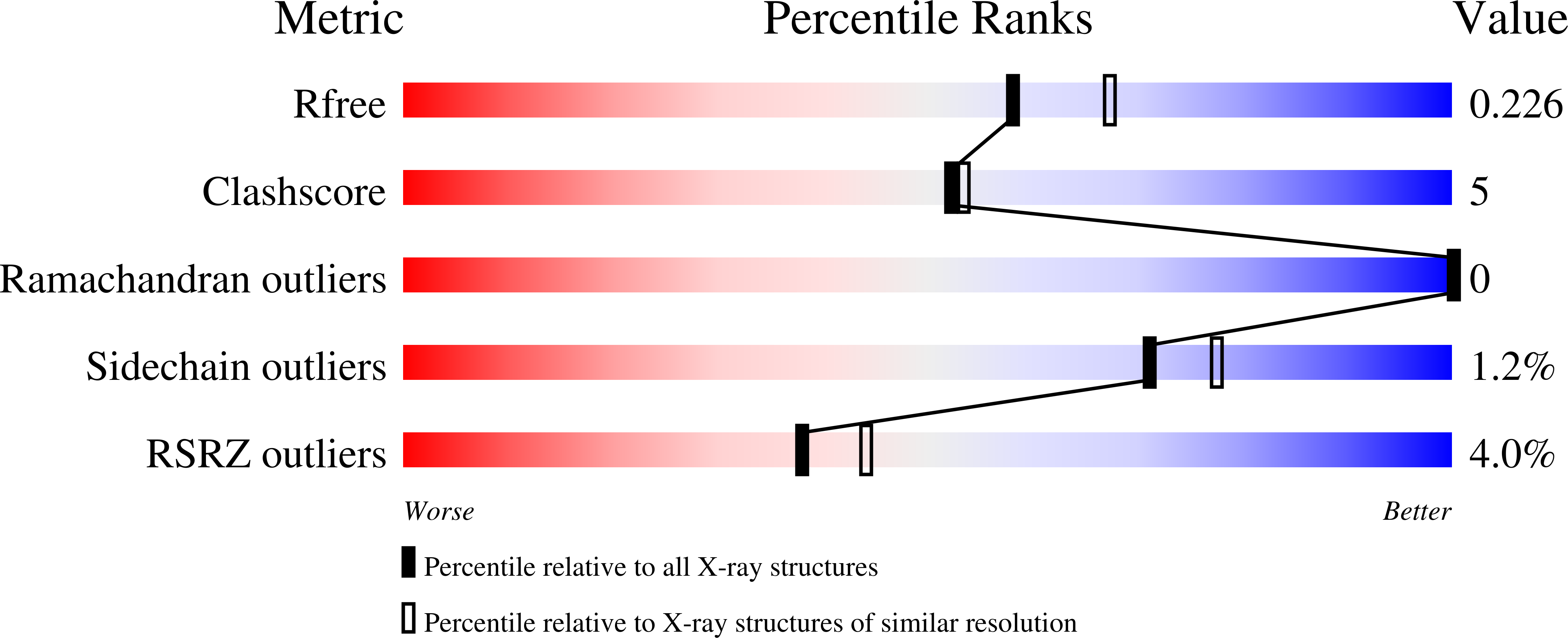
  • R-Value Free: 0.227 
  • R-Value Work: 0.191 
  • R-Value Observed: 0.192 

Starting Model: experimental
View more details

wwPDB Validation   3D Report Full Report


Ligand Structure Quality Assessment 


This is version 1.2 of the entry. See complete history


Literature

Millisecond cryo-trapping by the spitrobot crystal plunger simplifies time-resolved crystallography.

Mehrabi, P.Sung, S.von Stetten, D.Prester, A.Hatton, C.E.Kleine-Dopke, S.Berkes, A.Gore, G.Leimkohl, J.P.Schikora, H.Kollewe, M.Rohde, H.Wilmanns, M.Tellkamp, F.Schulz, E.C.

(2023) Nat Commun 14: 2365-2365

  • DOI: https://doi.org/10.1038/s41467-023-37834-w
  • Primary Citation of Related Structures:  
    8AW8, 8AW9, 8AWB, 8AWC, 8AWD, 8AWE, 8AWF, 8AWS, 8AWU, 8AWV, 8AWX, 8AWY, 8B03, 8B05, 8B06, 8B08, 8B2O, 8B2V, 8B2W, 8B3M

  • PubMed Abstract: 

    We introduce the spitrobot, a protein crystal plunger, enabling reaction quenching via cryo-trapping with a time-resolution in the millisecond range. Protein crystals are mounted on canonical micromeshes on an electropneumatic piston, where the crystals are kept in a humidity and temperature-controlled environment, then reactions are initiated via the liquid application method (LAMA) and plunging into liquid nitrogen is initiated after an electronically set delay time to cryo-trap intermediate states. High-magnification images are automatically recorded before and after droplet deposition, prior to plunging. The SPINE-standard sample holder is directly plunged into a storage puck, enabling compatibility with high-throughput infrastructure. Here we demonstrate binding of glucose and 2,3-butanediol in microcrystals of xylose isomerase, and of avibactam and ampicillin in microcrystals of the extended spectrum beta-lactamase CTX-M-14. We also trap reaction intermediates and conformational changes in macroscopic crystals of tryptophan synthase to demonstrate that the spitrobot enables insight into catalytic events.


  • Organizational Affiliation

    Institute for Nanostructure and Solid-State Physics, Universität Hamburg, Hamburg, Germany. pedram.mehrabi@uni-hamburg.de.


Macromolecules
Find similar proteins by:  (by identity cutoff)  |  3D Structure
Entity ID: 1
MoleculeChains Sequence LengthOrganismDetailsImage
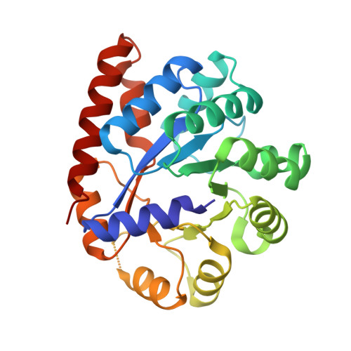
Tryptophan synthase alpha chain268Salmonella enterica subsp. enterica serovar TyphimuriumMutation(s): 0 
Gene Names: trpASTM1727
EC: 4.2.1.20
UniProt
Find proteins for P00929 (Salmonella typhimurium (strain LT2 / SGSC1412 / ATCC 700720))
Explore P00929 
Go to UniProtKB:  P00929
Entity Groups  
Sequence Clusters30% Identity50% Identity70% Identity90% Identity95% Identity100% Identity
UniProt GroupP00929
Sequence Annotations
Expand
  • Reference Sequence
Find similar proteins by:  (by identity cutoff)  |  3D Structure
Entity ID: 2
MoleculeChains Sequence LengthOrganismDetailsImage
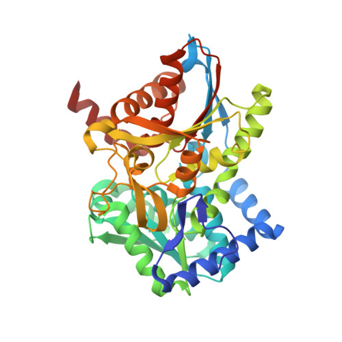
Tryptophan synthase beta chain397Salmonella enterica subsp. enterica serovar TyphimuriumMutation(s): 0 
Gene Names: trpBSTM1726
EC: 4.2.1.20
UniProt
Find proteins for P0A2K1 (Salmonella typhimurium (strain LT2 / SGSC1412 / ATCC 700720))
Explore P0A2K1 
Go to UniProtKB:  P0A2K1
Entity Groups  
Sequence Clusters30% Identity50% Identity70% Identity90% Identity95% Identity100% Identity
UniProt GroupP0A2K1
Sequence Annotations
Expand
  • Reference Sequence
Small Molecules
Ligands 4 Unique
IDChains Name / Formula / InChI Key2D Diagram3D Interactions
PLS (Subject of Investigation/LOI)
Query on PLS

Download Ideal Coordinates CCD File 
D [auth B][3-HYDROXY-2-METHYL-5-PHOSPHONOOXYMETHYL-PYRIDIN-4-YLMETHYL]-SERINE
C11 H17 N2 O8 P
ODVKKQWXKRZJLG-VIFPVBQESA-N
CS
Query on CS

Download Ideal Coordinates CCD File 
C [auth B]CESIUM ION
Cs
NCMHKCKGHRPLCM-UHFFFAOYSA-N
IND
Query on IND

Download Ideal Coordinates CCD File 
E [auth B]INDOLE
C8 H7 N
SIKJAQJRHWYJAI-UHFFFAOYSA-N
CL
Query on CL

Download Ideal Coordinates CCD File 
F [auth B],
G [auth B],
H [auth B]
CHLORIDE ION
Cl
VEXZGXHMUGYJMC-UHFFFAOYSA-M
Experimental Data & Validation

Experimental Data

  • Method: X-RAY DIFFRACTION
  • Resolution: 2.10 Å
  • R-Value Free: 0.227 
  • R-Value Work: 0.191 
  • R-Value Observed: 0.192 
  • Space Group: C 1 2 1
Unit Cell:
Length ( Å )Angle ( ˚ )
a = 180.99α = 90
b = 59.108β = 94.6
c = 66.848γ = 90
Software Package:
Software NamePurpose
XDSdata reduction
Aimlessdata scaling
PHENIXrefinement
PDB_EXTRACTdata extraction
PHASERphasing

Structure Validation

View Full Validation Report


Ligand Structure Quality Assessment 


Entry History & Funding Information

Deposition Data


Funding OrganizationLocationGrant Number
H2020 Marie Curie Actions of the European CommissionEuropean Union664726

Revision History  (Full details and data files)

  • Version 1.0: 2023-05-03
    Type: Initial release
  • Version 1.1: 2023-05-24
    Changes: Database references
  • Version 1.2: 2024-02-07
    Changes: Data collection, Refinement description