8EYU

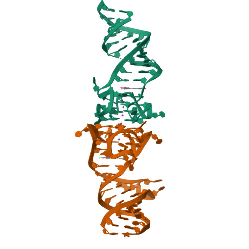
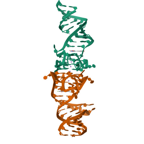
Structure of Beetroot dimer bound to DFAME


Experimental Data Snapshot

  • Method: X-RAY DIFFRACTION
  • Resolution: 1.95 Å
  • R-Value Free: 0.204 
  • R-Value Work: 0.178 
  • R-Value Observed: 0.181 

Starting Model: experimental
View more details

wwPDB Validation   3D Report Full Report


Ligand Structure Quality Assessment 


This is version 1.2 of the entry. See complete history


Literature

Co-crystal structures of the fluorogenic aptamer Beetroot show that close homology may not predict similar RNA architecture.

Passalacqua, L.F.M.Starich, M.R.Link, K.A.Wu, J.Knutson, J.R.Tjandra, N.Jaffrey, S.R.Ferre-D'Amare, A.R.

(2023) Nat Commun 14: 2969-2969

  • DOI: https://doi.org/10.1038/s41467-023-38683-3
  • Primary Citation of Related Structures:  
    8EYU, 8EYV, 8EYW, 8F0N

  • PubMed Abstract: 

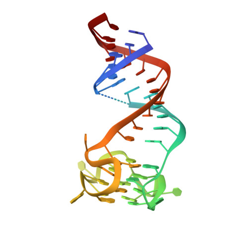
    Beetroot is a homodimeric in vitro selected RNA that binds and activates DFAME, a conditional fluorophore derived from GFP. It is 70% sequence-identical to the previously characterized homodimeric aptamer Corn, which binds one molecule of its cognate fluorophore DFHO at its interprotomer interface. We have now determined the Beetroot-DFAME co-crystal structure at 1.95 Å resolution, discovering that this RNA homodimer binds two molecules of the fluorophore, at sites separated by ~30 Å. In addition to this overall architectural difference, the local structures of the non-canonical, complex quadruplex cores of Beetroot and Corn are distinctly different, underscoring how subtle RNA sequence differences can give rise to unexpected structural divergence. Through structure-guided engineering, we generated a variant that has a 12-fold fluorescence activation selectivity switch toward DFHO. Beetroot and this variant form heterodimers and constitute the starting point for engineered tags whose through-space inter-fluorophore interaction could be used to monitor RNA dimerization.


  • Organizational Affiliation

    Biochemistry and Biophysics Center, National Heart, Lung, and Blood Institute, National Institutes of Health, Bethesda, MD, USA.


Macromolecules
Find similar nucleic acids by:  (by identity cutoff)  |  3D Structure
Entity ID: 1
MoleculeChains LengthOrganismImage
RNA (49-MER)A [auth B],
B [auth A]
49synthetic construct
Sequence Annotations
Expand
  • Reference Sequence
Small Molecules
Ligands 2 Unique
IDChains Name / Formula / InChI Key2D Diagram3D Interactions
X5R (Subject of Investigation/LOI)
Query on X5R

Download Ideal Coordinates CCD File 
E [auth B],
I [auth A]
methyl (2E)-3-{(4Z)-4-[(3,5-difluoro-4-hydroxyphenyl)methylidene]-1-methyl-5-oxo-4,5-dihydro-1H-imidazol-2-yl}prop-2-enoate
C15 H12 F2 N2 O4
LXQQJPJEYBZZSO-ZDVGJPJSSA-N
K
Query on K

Download Ideal Coordinates CCD File 
C [auth B],
D [auth B],
F [auth A],
G [auth A],
H [auth A]
POTASSIUM ION
K
NPYPAHLBTDXSSS-UHFFFAOYSA-N
Experimental Data & Validation

Experimental Data

  • Method: X-RAY DIFFRACTION
  • Resolution: 1.95 Å
  • R-Value Free: 0.204 
  • R-Value Work: 0.178 
  • R-Value Observed: 0.181 
  • Space Group: P 1 21 1
Unit Cell:
Length ( Å )Angle ( ˚ )
a = 29.937α = 90
b = 50.742β = 94.083
c = 95.555γ = 90
Software Package:
Software NamePurpose
PHENIXrefinement
Cootmodel building
xia2data reduction
DIALSdata scaling
PHASERphasing

Structure Validation

View Full Validation Report


Ligand Structure Quality Assessment 


Entry History & Funding Information

Deposition Data


Funding OrganizationLocationGrant Number
National Institutes of Health/National Heart, Lung, and Blood Institute (NIH/NHLBI)United States--

Revision History  (Full details and data files)

  • Version 1.0: 2023-05-31
    Type: Initial release
  • Version 1.1: 2023-09-20
    Changes: Data collection, Database references
  • Version 1.2: 2023-10-25
    Changes: Refinement description