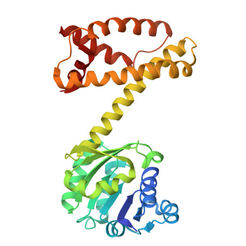
8P2J

Imine Reductase from Ajellomyces dermatitidis in space group C21


Experimental Data Snapshot

  • Method: X-RAY DIFFRACTION
  • Resolution: 1.73 Å
  • R-Value Free: 0.193 
  • R-Value Work: 0.166 
  • R-Value Observed: 0.168 

Starting Model: experimental
View more details

wwPDB Validation   3D Report Full Report


Ligand Structure Quality Assessment 


This is version 1.1 of the entry. See complete history


Literature

Structure of the imine reductase from Ajellomyces dermatitidis in three crystal forms.

Sharma, M.Cuetos, A.Willliams, A.Gonzalez-Martinez, D.Grogan, G.

(2023) Acta Crystallogr F Struct Biol Commun 79: 224-230

  • DOI: https://doi.org/10.1107/S2053230X23006672
  • Primary Citation of Related Structures:  
    8OZV, 8OZW, 8P2J

  • PubMed Abstract: 

    The NADPH-dependent imine reductase from Ajellomyces dermatitidis (AdRedAm) catalyzes the reductive amination of certain ketones with amine donors supplied in an equimolar ratio. The structure of AdRedAm has been determined in three forms. The first form, which belongs to space group P3 1 21 and was refined to 2.01 Å resolution, features two molecules (one dimer) in the asymmetric unit in complex with the redox-inactive cofactor NADPH 4 . The second form, which belongs to space group C2 1 and was refined to 1.73 Å resolution, has nine molecules (four and a half dimers) in the asymmetric unit, each complexed with NADP + . The third form, which belongs to space group P3 1 21 and was refined to 1.52 Å resolution, has one molecule (one half-dimer) in the asymmetric unit. This structure was again complexed with NADP + and also with the substrate 2,2-difluoroacetophenone. The different data sets permit the analysis of AdRedAm in different conformational states and also reveal the molecular basis of stereoselectivity in the transformation of fluorinated acetophenone substrates by the enzyme.


  • Organizational Affiliation

    Department of Chemistry, University of York, Heslington, York YO10 5DD, United Kingdom.


Macromolecules
Find similar proteins by:  (by identity cutoff)  |  3D Structure
Entity ID: 1
MoleculeChains Sequence LengthOrganismDetailsImage
Oxidoreductase
A, B, C, D, E
A, B, C, D, E, F, G, H, I
294Blastomyces dermatitidisMutation(s): 1 
Gene Names: BDBG_03427
UniProt
Find proteins for A0A179UH34 (Blastomyces gilchristii (strain SLH14081))
Explore A0A179UH34 
Go to UniProtKB:  A0A179UH34
Entity Groups  
Sequence Clusters30% Identity50% Identity70% Identity90% Identity95% Identity100% Identity
UniProt GroupA0A179UH34
Sequence Annotations
Expand
  • Reference Sequence
Small Molecules
Ligands 1 Unique
IDChains Name / Formula / InChI Key2D Diagram3D Interactions
NDP (Subject of Investigation/LOI)
Query on NDP

Download Ideal Coordinates CCD File 
J [auth A]
K [auth B]
L [auth C]
M [auth D]
N [auth E]
J [auth A],
K [auth B],
L [auth C],
M [auth D],
N [auth E],
O [auth F],
P [auth G],
Q [auth H],
R [auth I]
NADPH DIHYDRO-NICOTINAMIDE-ADENINE-DINUCLEOTIDE PHOSPHATE
C21 H30 N7 O17 P3
ACFIXJIJDZMPPO-NNYOXOHSSA-N
Experimental Data & Validation

Experimental Data

  • Method: X-RAY DIFFRACTION
  • Resolution: 1.73 Å
  • R-Value Free: 0.193 
  • R-Value Work: 0.166 
  • R-Value Observed: 0.168 
  • Space Group: C 1 2 1
Unit Cell:
Length ( Å )Angle ( ˚ )
a = 204.62α = 90
b = 87.97β = 108.45
c = 162.58γ = 90
Software Package:
Software NamePurpose
REFMACrefinement
XDSdata reduction
SCALAdata scaling
MOLREPphasing

Structure Validation

View Full Validation Report


Ligand Structure Quality Assessment 


Entry History & Funding Information

Deposition Data


Funding OrganizationLocationGrant Number
Biotechnology and Biological Sciences Research Council (BBSRC)United KingdomBB/M006832/1

Revision History  (Full details and data files)

  • Version 1.0: 2023-08-30
    Type: Initial release
  • Version 1.1: 2023-09-13
    Changes: Database references