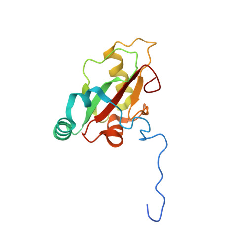
8S1M | pdb_00008s1m

Crystal structure of human GABARAP fused to EGFR (1076-1099)


Experimental Data Snapshot

  • Method: X-RAY DIFFRACTION
  • Resolution: 2.05 Å
  • R-Value Free: 
    0.230 (Depositor) 
  • R-Value Work: 
    0.203 (Depositor) 
  • R-Value Observed: 
    0.205 (Depositor) 

Starting Model: experimental
View more details

wwPDB Validation   3D Report Full Report


This is version 1.1 of the entry. See complete history


Literature

GABARAP interacts with EGFR - supporting the unique role of this hAtg8 protein during receptor trafficking.

Uffing, A.Weiergraber, O.H.Schwarten, M.Hoffmann, S.Willbold, D.

(2024) FEBS Lett 598: 2656-2669

  • DOI: https://doi.org/10.1002/1873-3468.14997
  • Primary Citation of Related Structures:  
    8S1M

  • PubMed Abstract: 

    The human Atg8 family member GABARAP is involved in numerous autophagy-related and -unrelated processes. We recently observed that specifically the deficiency of GABARAP enhances epidermal growth factor receptor (EGFR) degradation upon ligand stimulation. Here, we report on two putative LC3-interacting regions (LIRs) within EGFR, the first of which (LIR1) is selected as a GABARAP binding site in silico. Indeed, in vitro interaction studies reveal preferential binding of LIR1 to GABARAP and GABARAPL1. Our X-ray data demonstrate interaction of core LIR1 residues FLPV with both hydrophobic pockets of GABARAP suggesting canonical binding. Although LIR1 occupies the LIR docking site, GABARAP Y49 and L50 appear dispensable in this case. Our data support the hypothesis that GABARAP affects the fate of EGFR at least in part through direct binding.


  • Organizational Affiliation

    Heinrich-Heine-Universität Düsseldorf, Mathematisch-Naturwissenschaftliche Fakultät, Institut für Physikalische Biologie, Düsseldorf, Germany.


Macromolecules
Find similar proteins by:  (by identity cutoff)  |  3D Structure
Entity ID: 1
MoleculeChains Sequence LengthOrganismDetailsImage
Epidermal growth factor receptor,Gamma-aminobutyric acid receptor-associated protein145Homo sapiensMutation(s): 0 
Gene Names: EGFRERBBERBB1HER1GABARAPFLC3BHT004
EC: 2.7.10.1
UniProt & NIH Common Fund Data Resources
Find proteins for P00533 (Homo sapiens)
Explore P00533 
Go to UniProtKB:  P00533
PHAROS:  P00533
GTEx:  ENSG00000146648 
Find proteins for O95166 (Homo sapiens)
Explore O95166 
Go to UniProtKB:  O95166
PHAROS:  O95166
GTEx:  ENSG00000170296 
Entity Groups  
Sequence Clusters30% Identity50% Identity70% Identity90% Identity95% Identity100% Identity
UniProt GroupsP00533O95166
Sequence Annotations
Expand
  • Reference Sequence
Small Molecules
Ligands 2 Unique
IDChains Name / Formula / InChI Key2D Diagram3D Interactions
GOL
Query on GOL

Download Ideal Coordinates CCD File 
C [auth A]GLYCEROL
C3 H8 O3
PEDCQBHIVMGVHV-UHFFFAOYSA-N
CL
Query on CL

Download Ideal Coordinates CCD File 
B [auth A]CHLORIDE ION
Cl
VEXZGXHMUGYJMC-UHFFFAOYSA-M
Experimental Data & Validation

Experimental Data

  • Method: X-RAY DIFFRACTION
  • Resolution: 2.05 Å
  • R-Value Free:  0.230 (Depositor) 
  • R-Value Work:  0.203 (Depositor) 
  • R-Value Observed: 0.205 (Depositor) 
Space Group: I 2 3
Unit Cell:
Length ( Å )Angle ( ˚ )
a = 101.28α = 90
b = 101.28β = 90
c = 101.28γ = 90
Software Package:
Software NamePurpose
PHENIXrefinement
XDSdata reduction
XDSdata scaling
MOLREPphasing

Structure Validation

View Full Validation Report


Entry History & Funding Information

Deposition Data


Funding OrganizationLocationGrant Number
German Research Foundation (DFG)Germany267205415

Revision History  (Full details and data files)

  • Version 1.0: 2024-08-14
    Type: Initial release
  • Version 1.1: 2025-01-15
    Changes: Database references, Structure summary