8S39

Crystal structure of Medicago truncatula glutamate dehydrogenase 2 in complex with isophthalic acid and NAD


Experimental Data Snapshot

  • Method: X-RAY DIFFRACTION
  • Resolution: 2.00 Å
  • R-Value Free: 
    0.212 (Depositor), 0.212 (DCC) 
  • R-Value Work: 
    0.163 (Depositor), 0.170 (DCC) 
  • R-Value Observed: 
    0.163 (Depositor) 

Starting Model: experimental
View more details

wwPDB Validation   3D Report Full Report


Ligand Structure Quality Assessment 

Created with Raphaël 2.3.0Worse 01 BetterLigand structure goodness of fit to experimental dataBest fitted NADClick on this verticalbar to view detailsBest fitted 8G0Click on this verticalbar to view details

This is version 1.0 of the entry. See complete history


Literature

Legume-type glutamate dehydrogenase: Structure, activity, and inhibition studies.

Grzechowiak, M.Sliwiak, J.Link, A.Ruszkowski, M.

(2024) Int J Biol Macromol 278: 134648-134648

  • DOI: https://doi.org/10.1016/j.ijbiomac.2024.134648
  • Primary Citation of Related Structures:  
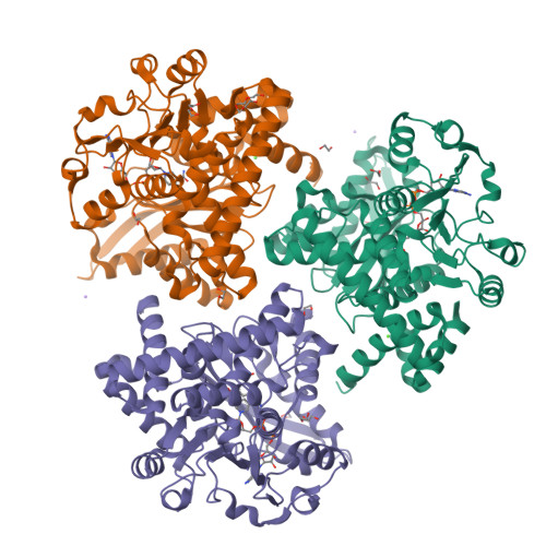
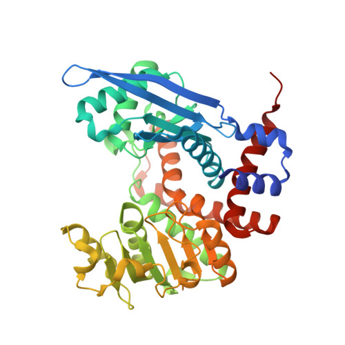
    8S38, 8S39, 8S3A, 8S3B, 8S3C, 8S3D

  • PubMed Abstract: 

    Glutamate dehydrogenases (GDHs) are key enzymes at the crossroads of N and C metabolism in plants. Legumes, whose N metabolism is particularly intricate, possess a unique type of GDH. This study presents an analysis of a legume-type GDH (isoform 2) from Medicago truncatula (MtGDH2). We measured MtGDH2 activity in both the Glu → 2-oxoglutarate (2OG) and 2OG → Glu reaction directions and obtained kinetic parameters for Glu, 2OG, NAD + , and NADH. Inhibition assays revealed that compounds possessing di- or tricarboxylates act as inhibitors of plant GDHs. Interestingly, 2,6-pyridinedicarboxylate (PYR) weakly inhibits MtGDH2 compared to Arabidopsis thaliana homologs. Furthermore, we explored tetrazole derivatives to discover 3-(1H-tetrazol-5-yl)benzoic acid (TBA) as an MtGDH2 inhibitor. The kinetic experiments are supported by six crystal structures, solved as: (i) unliganded enzyme, (ii) trapping the reaction intermediate 2-amino-2-hydroxyglutarate and NAD + , and also complexed with NAD + and inhibitors such as (iii) citrate, (iv) PYR, (v) isophthalate, and (vi) TBA. The complex with TBA revealed a new mode of action that, in contrast to other inhibitors, prevents domain closure. This discovery points to TBA as a starting point for the development of novel GDH inhibitors to study the functions of GDH in plants and potentially boost biomass production.


  • Organizational Affiliation

    Department of Structural Biology of Eukaryotes, Institute of Bioorganic Chemistry, Polish Academy of Sciences, Poznan 61-704, Poland.


Macromolecules
Find similar proteins by:  (by identity cutoff)  |  3D Structure
Entity ID: 1
MoleculeChains Sequence LengthOrganismDetailsImage
Glutamate dehydrogenase
A, B, C
414Medicago truncatulaMutation(s): 0 
Gene Names: 11433210MTR_5g013470MtrunA17_Chr5g0399821
UniProt
Find proteins for G7JYL4 (Medicago truncatula)
Explore G7JYL4 
Go to UniProtKB:  G7JYL4
Entity Groups  
Sequence Clusters30% Identity50% Identity70% Identity90% Identity95% Identity100% Identity
UniProt GroupG7JYL4
Sequence Annotations
Expand
  • Reference Sequence
Small Molecules
Ligands 7 Unique
IDChains Name / Formula / InChI Key2D Diagram3D Interactions
NAD (Subject of Investigation/LOI)
Query on NAD

Download Ideal Coordinates CCD File 
G [auth A],
O [auth B],
W [auth C]
NICOTINAMIDE-ADENINE-DINUCLEOTIDE
C21 H27 N7 O14 P2
BAWFJGJZGIEFAR-NNYOXOHSSA-N
PG4
Query on PG4

Download Ideal Coordinates CCD File 
L [auth B]TETRAETHYLENE GLYCOL
C8 H18 O5
UWHCKJMYHZGTIT-UHFFFAOYSA-N
8G0 (Subject of Investigation/LOI)
Query on 8G0

Download Ideal Coordinates CCD File 
X [auth C]benzene-1,3-dicarboxylic acid
C8 H6 O4
QQVIHTHCMHWDBS-UHFFFAOYSA-N
PEG
Query on PEG

Download Ideal Coordinates CCD File 
J [auth B]DI(HYDROXYETHYL)ETHER
C4 H10 O3
MTHSVFCYNBDYFN-UHFFFAOYSA-N
EDO
Query on EDO

Download Ideal Coordinates CCD File 
D [auth A]
E [auth A]
F [auth A]
K [auth B]
M [auth B]
1,2-ETHANEDIOL
C2 H6 O2
LYCAIKOWRPUZTN-UHFFFAOYSA-N
CA
Query on CA

Download Ideal Coordinates CCD File 
I [auth A],
Q [auth B],
R [auth B]
CALCIUM ION
Ca
BHPQYMZQTOCNFJ-UHFFFAOYSA-N
NA
Query on NA

Download Ideal Coordinates CCD File 
H [auth A],
P [auth B]
SODIUM ION
Na
FKNQFGJONOIPTF-UHFFFAOYSA-N
Experimental Data & Validation

Experimental Data

  • Method: X-RAY DIFFRACTION
  • Resolution: 2.00 Å
  • R-Value Free:  0.212 (Depositor), 0.212 (DCC) 
  • R-Value Work:  0.163 (Depositor), 0.170 (DCC) 
  • R-Value Observed: 0.163 (Depositor) 
Space Group: I 21 21 21
Unit Cell:
Length ( Å )Angle ( ˚ )
a = 94.728α = 90
b = 162.769β = 90
c = 216.988γ = 90
Software Package:
Software NamePurpose
PHENIXrefinement
XDSdata reduction
XSCALEdata scaling
PHASERphasing

Structure Validation

View Full Validation Report


Ligand Structure Quality Assessment 

Created with Raphaël 2.3.0Worse 01 BetterLigand structure goodness of fit to experimental dataBest fitted NADClick on this verticalbar to view detailsBest fitted 8G0Click on this verticalbar to view details

Entry History & Funding Information

Deposition Data


Funding OrganizationLocationGrant Number
Polish National Science CentrePolandSONATA 2018/31/D/NZ1/03630

Revision History  (Full details and data files)

  • Version 1.0: 2024-09-04
    Type: Initial release