1RWN | pdb_00001rwn

Crystal structure of human caspase-1 in complex with 3-{2-ethyl-6-[4-(quinoxalin-2-ylamino)-benzoylamino]-hexanoylamino}-4-oxo-butyric acid


Experimental Data Snapshot

  • Method: X-RAY DIFFRACTION
  • Resolution: 2.00 Å
  • R-Value Free: 
    0.236 (Depositor), 0.240 (DCC) 
  • R-Value Work: 
    0.201 (Depositor), 0.200 (DCC) 
  • R-Value Observed: 
    0.203 (Depositor) 

Starting Model: experimental
View more details

wwPDB Validation   3D Report Full Report


Ligand Structure Quality Assessment 

Created with Raphaël 2.3.0Worse 01 BetterLigand structure goodness of fit to experimental dataBest fitted 4QBClick on this verticalbar to view details

This is version 1.4 of the entry. See complete history


Literature

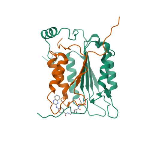
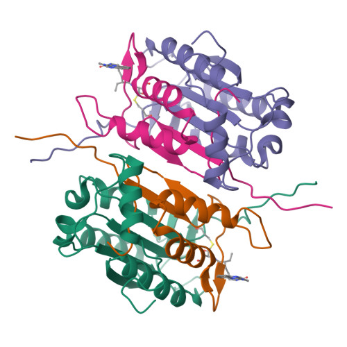
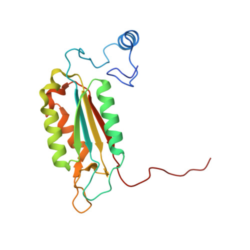
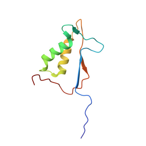
Structural analysis of caspase-1 inhibitors derived from Tethering.

O'Brien, T.Fahr, B.T.Sopko, M.M.Lam, J.W.Waal, N.D.Raimundo, B.C.Purkey, H.E.Pham, P.Romanowski, M.J.

(2005) Acta Crystallogr Sect F Struct Biol Cryst Commun 61: 451-458

  • DOI: https://doi.org/10.1107/S1744309105010109
  • Primary Citation of Related Structures:  
    1RWK, 1RWM, 1RWN, 1RWO, 1RWP

  • PubMed Abstract: 

    Caspase-1 is a key endopeptidase responsible for the post-translational processing of the IL-1beta and IL-18 cytokines and small-molecule inhibitors that modulate the activity of this enzyme are predicted to be important therapeutic treatments for many inflammatory diseases. A fragment-assembly approach, accompanied by structural analysis, was employed to generate caspase-1 inhibitors. With the aid of Tethering with extenders (small molecules that bind to the active-site cysteine and contain a free thiol), two novel fragments that bound to the active site and made a disulfide bond with the extender were identified by mass spectrometry. Direct linking of each fragment to the extender generated submicromolar reversible inhibitors that significantly reduced secretion of IL-1beta but not IL-6 from human peripheral blood mononuclear cells. Thus, Tethering with extenders facilitated rapid identification and synthesis of caspase-1 inhibitors with cell-based activity and subsequent structural analyses provided insights into the enzyme's ability to accommodate different inhibitor-binding modes in the active site.


  • Organizational Affiliation

    Department of Biology, Sunesis Pharmaceuticals Inc., USA.


Macromolecules
Find similar proteins by:  (by identity cutoff)  |  3D Structure
Entity ID: 1
MoleculeChains Sequence LengthOrganismDetailsImage
Interleukin-1 beta convertase178Homo sapiensMutation(s): 0 
Gene Names: CASP1IL1BCIL1BCE
EC: 3.4.22.36
UniProt & NIH Common Fund Data Resources
Find proteins for P29466 (Homo sapiens)
Explore P29466 
Go to UniProtKB:  P29466
PHAROS:  P29466
GTEx:  ENSG00000137752 
Entity Groups  
Sequence Clusters30% Identity50% Identity70% Identity90% Identity95% Identity100% Identity
UniProt GroupP29466
Sequence Annotations
Expand
  • Reference Sequence
Find similar proteins by:  (by identity cutoff)  |  3D Structure
Entity ID: 2
MoleculeChains Sequence LengthOrganismDetailsImage
Interleukin-1 beta convertase88Homo sapiensMutation(s): 0 
Gene Names: CASP1IL1BCIL1BCE
EC: 3.4.22.36
UniProt & NIH Common Fund Data Resources
Find proteins for P29466 (Homo sapiens)
Explore P29466 
Go to UniProtKB:  P29466
PHAROS:  P29466
GTEx:  ENSG00000137752 
Entity Groups  
Sequence Clusters30% Identity50% Identity70% Identity90% Identity95% Identity100% Identity
UniProt GroupP29466
Sequence Annotations
Expand
  • Reference Sequence
Small Molecules
Ligands 1 Unique
IDChains Name / Formula / InChI Key2D Diagram3D Interactions
4QB
Query on 4QB

Download Ideal Coordinates CCD File 
C [auth A]3-{2-ETHYL-6-[4-(QUINOXALIN-2-YLAMINO)-BENZOYLAMINO]-HEXANOYLAMINO}-4-OXO-BUTYRIC ACID
C27 H31 N5 O5
LFXGHNVCJJFHJJ-NQIIRXRSSA-N
Experimental Data & Validation

Experimental Data

  • Method: X-RAY DIFFRACTION
  • Resolution: 2.00 Å
  • R-Value Free:  0.236 (Depositor), 0.240 (DCC) 
  • R-Value Work:  0.201 (Depositor), 0.200 (DCC) 
  • R-Value Observed: 0.203 (Depositor) 
Space Group: P 43 21 2
Unit Cell:
Length ( Å )Angle ( ˚ )
a = 63.147α = 90
b = 63.147β = 90
c = 161.771γ = 90
Software Package:
Software NamePurpose
REFMACrefinement
MOSFLMdata reduction
d*TREKdata scaling
AMoREphasing

Structure Validation

View Full Validation Report


Ligand Structure Quality Assessment 

Created with Raphaël 2.3.0Worse 01 BetterLigand structure goodness of fit to experimental dataBest fitted 4QBClick on this verticalbar to view details

Entry History 

Deposition Data

Revision History  (Full details and data files)

  • Version 1.0: 2004-12-28
    Type: Initial release
  • Version 1.1: 2008-04-29
    Changes: Version format compliance
  • Version 1.2: 2011-07-13
    Changes: Derived calculations, Version format compliance
  • Version 1.3: 2023-08-23
    Changes: Data collection, Database references, Derived calculations, Refinement description
  • Version 1.4: 2024-10-30
    Changes: Structure summary