A novel, potent dual inhibitor of the leukocyte proteases cathepsin G and chymase: molecular mechanisms and anti-inflammatory activity in vivo.
de Garavilla, L., Greco, M.N., Sukumar, N., Chen, Z.W., Pineda, A.O., Mathews, F.S., Di Cera, E., Giardino, E.C., Wells, G.I., Haertlein, B.J., Kauffman, J.A., Corcoran, T.W., Derian, C.K., Eckardt, A.J., Damiano, B.P., Andrade-Gordon, P., Maryanoff, B.E.(2005) J Biological Chem 280: 18001-18007
- PubMed: 15741158
- DOI: https://doi.org/10.1074/jbc.M501302200
- Primary Citation of Related Structures:
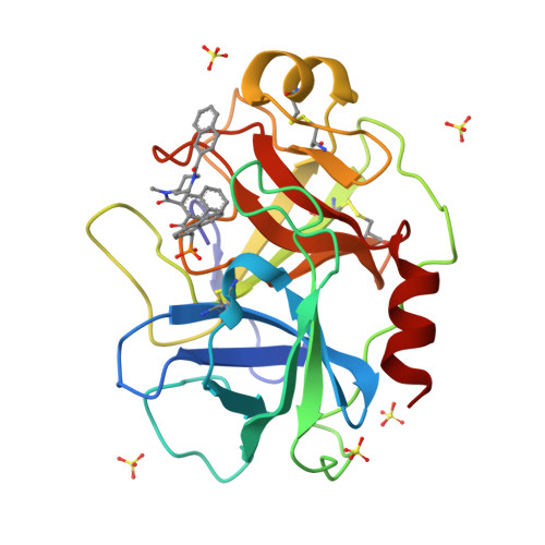
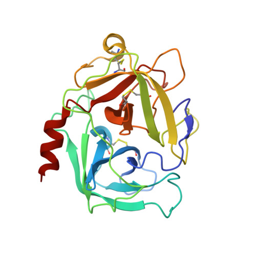
1T31, 1T32 - PubMed Abstract:
Certain leukocytes release serine proteases that sustain inflammatory processes and cause disease conditions, such as asthma and chronic obstructive pulmonary disease. We identified beta-ketophosphonate 1 (JNJ-10311795; RWJ-355871) as a novel, potent dual inhibitor of neutrophil cathepsin G (K(i) = 38 nm) and mast cell chymase (K(i) = 2.3 nm). The x-ray crystal structures of 1 complexed with human cathepsin G (1.85 A) and human chymase (1.90 A) reveal the molecular basis of the dual inhibition. Ligand 1 occupies the S(1) and S(2) subsites of cathepsin G and chymase similarly, with the 2-naphthyl in S(1), the 1-naphthyl in S(2), and the phosphonate group in a complex network of hydrogen bonds. Surprisingly, however, the carboxamido-N-(naphthalene-2-carboxyl)piperidine group is found to bind in two distinct conformations. In cathepsin G, this group occupies the hydrophobic S(3)/S(4) subsites, whereas in chymase, it does not; rather, it folds onto the 1-naphthyl group of the inhibitor itself. Compound 1 exhibited noteworthy anti-inflammatory activity in rats for glycogen-induced peritonitis and lipopolysaccharide-induced airway inflammation. In addition to a marked reduction in neutrophil influx, 1 reversed increases in inflammatory mediators interleukin-1alpha, interleukin-1beta, tissue necrosis factor-alpha, and monocyte chemotactic protein-1 in the glycogen model and reversed increases in airway nitric oxide levels in the lipopolysaccharide model. These findings demonstrate that it is possible to inhibit both cathepsin G and chymase with a single molecule and suggest an exciting opportunity in the treatment of asthma and chronic obstructive pulmonary disease.
Organizational Affiliation:
Drug Discovery, Johnson & Johnson Pharmaceutical Research and Development, Spring House, Pennsylvania 19477-0776, USA.