2HCB | pdb_00002hcb

Structure of AMPPCP-bound DnaA from Aquifex aeolicus


Experimental Data Snapshot

  • Method: X-RAY DIFFRACTION
  • Resolution: 3.51 Å
  • R-Value Free: 
    0.298 (Depositor), 0.250 (DCC) 
  • R-Value Work: 
    0.266 (Depositor), 0.250 (DCC) 
  • R-Value Observed: 
    0.267 (Depositor) 

Starting Model: experimental
View more details

wwPDB Validation   3D Report Full Report


Ligand Structure Quality Assessment 

Created with Raphaël 2.3.0Worse 01 BetterLigand structure goodness of fit to experimental dataBest fitted ACPClick on this verticalbar to view details

This is version 1.3 of the entry. See complete history


Literature

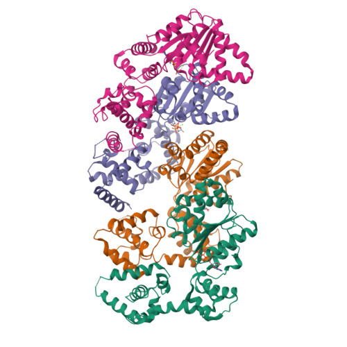
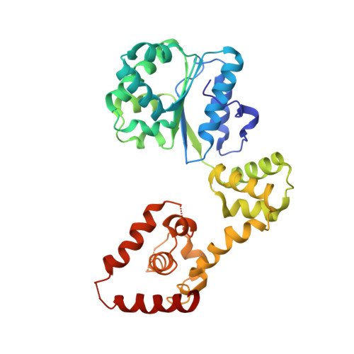
Structural basis for ATP-dependent DnaA assembly and replication-origin remodeling.

Erzberger, J.P.Mott, M.L.Berger, J.M.

(2006) Nat Struct Mol Biol 13: 676-683

  • DOI: https://doi.org/10.1038/nsmb1115

  • PubMed Abstract: 

    In bacteria, the initiation of replication is controlled by DnaA, a member of the ATPases associated with various cellular activities (AAA+) protein superfamily. ATP binding allows DnaA to transition from a monomeric state into a large oligomeric complex that remodels replication origins, triggers duplex melting and facilitates replisome assembly. The crystal structure of AMP-PCP-bound DnaA reveals a right-handed superhelix defined by specific protein-ATP interactions. The observed quaternary structure of DnaA, along with topology footprint assays, indicates that a right-handed DNA wrap is formed around the initiation nucleoprotein complex. This model clarifies how DnaA engages and unwinds bacterial origins and suggests that additional, regulatory AAA+ proteins engage DnaA at filament ends. Eukaryotic and archaeal initiators also have the structural elements that promote open-helix formation, indicating that a spiral, open-ring AAA+ assembly forms the core element of initiators in all domains of life.


  • Organizational Affiliation

    Division of Biochemistry and Molecular Biology, Molecular and Cell Biology Department, 327B Hildebrand Hall #3206, University of California, Berkeley, California 94720, USA.


Macromolecules
Find similar proteins by:  (by identity cutoff)  |  3D Structure
Entity ID: 1
MoleculeChains Sequence LengthOrganismDetailsImage
Chromosomal replication initiator protein dnaA
A, B, C, D
323Aquifex aeolicusMutation(s): 0 
Gene Names: dnaA
UniProt
Find proteins for O66659 (Aquifex aeolicus (strain VF5))
Explore O66659 
Go to UniProtKB:  O66659
Entity Groups  
Sequence Clusters30% Identity50% Identity70% Identity90% Identity95% Identity100% Identity
UniProt GroupO66659
Sequence Annotations
Expand
  • Reference Sequence
Small Molecules
Ligands 2 Unique
IDChains Name / Formula / InChI Key2D Diagram3D Interactions
ACP
Query on ACP

Download Ideal Coordinates CCD File 
F [auth A],
H [auth B],
J [auth C],
L [auth D]
PHOSPHOMETHYLPHOSPHONIC ACID ADENYLATE ESTER
C11 H18 N5 O12 P3
UFZTZBNSLXELAL-IOSLPCCCSA-N
MG
Query on MG

Download Ideal Coordinates CCD File 
E [auth A],
G [auth B],
I [auth C],
K [auth D]
MAGNESIUM ION
Mg
JLVVSXFLKOJNIY-UHFFFAOYSA-N
Experimental Data & Validation

Experimental Data

  • Method: X-RAY DIFFRACTION
  • Resolution: 3.51 Å
  • R-Value Free:  0.298 (Depositor), 0.250 (DCC) 
  • R-Value Work:  0.266 (Depositor), 0.250 (DCC) 
  • R-Value Observed: 0.267 (Depositor) 
Space Group: P 21 21 21
Unit Cell:
Length ( Å )Angle ( ˚ )
a = 99.076α = 90
b = 117.714β = 90
c = 200.994γ = 90
Software Package:
Software NamePurpose
REFMACrefinement
PDB_EXTRACTdata extraction
ADSCdata collection
HKL-2000data scaling
PHASERphasing

Structure Validation

View Full Validation Report


Ligand Structure Quality Assessment 

Created with Raphaël 2.3.0Worse 01 BetterLigand structure goodness of fit to experimental dataBest fitted ACPClick on this verticalbar to view details

Entry History 

Deposition Data

Revision History  (Full details and data files)

  • Version 1.0: 2006-07-25
    Type: Initial release
  • Version 1.1: 2008-05-01
    Changes: Version format compliance
  • Version 1.2: 2011-07-13
    Changes: Advisory, Version format compliance
  • Version 1.3: 2023-08-30
    Changes: Data collection, Database references, Derived calculations, Refinement description