3HQE | pdb_00003hqe

Crystal Structure of the decamer CGGGCGCCCG forming a Holliday junction


Experimental Data Snapshot

  • Method: X-RAY DIFFRACTION
  • Resolution: 2.94 Å
  • R-Value Free: 
    0.239 (Depositor), 0.230 (DCC) 
  • R-Value Work: 
    0.215 (Depositor), 0.210 (DCC) 
  • R-Value Observed: 
    0.217 (Depositor) 

Starting Model: experimental
View more details

wwPDB Validation   3D Report Full Report


This is version 1.2 of the entry. See complete history


Literature

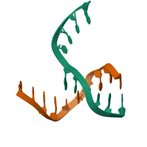
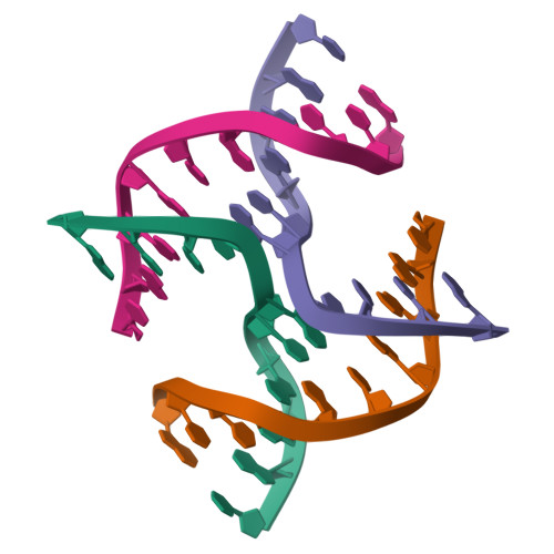
Sequence dependent structural variation of Holliday junction - crystal structure of d(CGGGCGCCCG)4

Venkadesh, S.Mandal, P.K.Gautham, N.

To be published.

Macromolecules

Find similar nucleic acids by:  Sequence   |   3D Structure  

Entity ID: 1
MoleculeChains LengthOrganismImage
5'-D(*CP*GP*GP*GP*CP*GP*CP*CP*CP*G)-3'
A, B
10N/A
Sequence Annotations
Expand
  • Reference Sequence
Experimental Data & Validation

Experimental Data

  • Method: X-RAY DIFFRACTION
  • Resolution: 2.94 Å
  • R-Value Free:  0.239 (Depositor), 0.230 (DCC) 
  • R-Value Work:  0.215 (Depositor), 0.210 (DCC) 
  • R-Value Observed: 0.217 (Depositor) 
Space Group: C 1 2 1
Unit Cell:
Length ( Å )Angle ( ˚ )
a = 65.004α = 90
b = 22.813β = 116.75
c = 40.708γ = 90
Software Package:
Software NamePurpose
MAR345dtbdata collection
AMoREphasing
REFMACrefinement
AUTOMARdata reduction
SCALEPACKdata scaling

Structure Validation

View Full Validation Report


Entry History 

Deposition Data

Revision History  (Full details and data files)

  • Version 1.0: 2010-06-09
    Type: Initial release
  • Version 1.1: 2011-07-13
    Changes: Version format compliance
  • Version 1.2: 2023-11-01
    Changes: Data collection, Database references, Refinement description