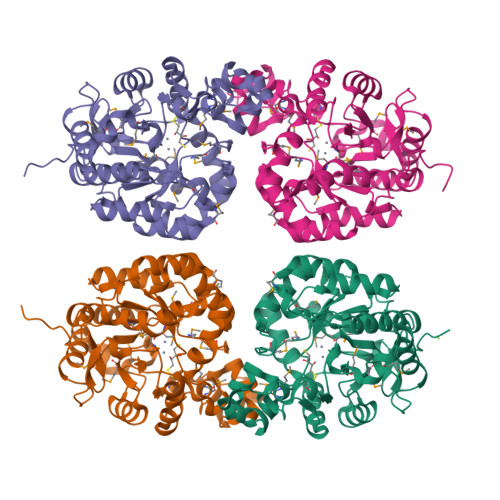
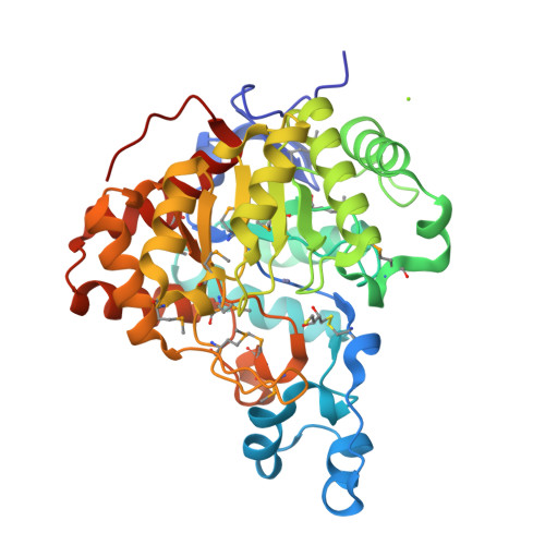
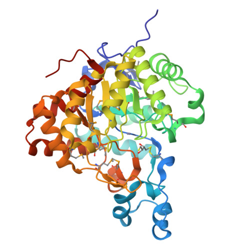
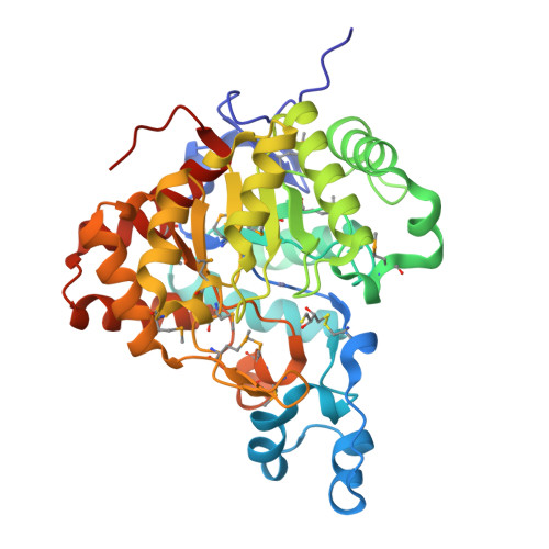
Crystal structure of a Resiniferatoxin-binding protein from Rhodobacter sphaeroides
Kumaran, D., Burley, S.K., Swaminathan, S.To be published.
Experimental Data Snapshot
Entity ID: 1 | |||||
|---|---|---|---|---|---|
| Molecule | Chains | Sequence Length | Organism | Details | Image |
| Resiniferatoxin-binding, phosphotriesterase-related protein | 364 | Cereibacter sphaeroides 2.4.1 | Mutation(s): 0 Gene Names: RHOS4_37320, RHOS4_37320 (RSP_3690), RSP_3690 |  | |
UniProt | |||||
Find proteins for Q3IVY4 (Cereibacter sphaeroides (strain ATCC 17023 / DSM 158 / JCM 6121 / CCUG 31486 / LMG 2827 / NBRC 12203 / NCIMB 8253 / ATH 2.4.1.)) Explore Q3IVY4 Go to UniProtKB: Q3IVY4 | |||||
Entity Groups | |||||
| Sequence Clusters | 30% Identity50% Identity70% Identity90% Identity95% Identity100% Identity | ||||
| UniProt Group | Q3IVY4 | ||||
Sequence AnnotationsExpand | |||||
| |||||
| Ligands 3 Unique | |||||
|---|---|---|---|---|---|
| ID | Chains | Name / Formula / InChI Key | 2D Diagram | 3D Interactions | |
| DTV Query on DTV | H [auth A], K [auth B], N [auth C], Q [auth D] | (2S,3S)-1,4-DIMERCAPTOBUTANE-2,3-DIOL C4 H10 O2 S2 VHJLVAABSRFDPM-QWWZWVQMSA-N |  | ||
| ZN Query on ZN | E [auth A] F [auth A] I [auth B] J [auth B] L [auth C] | ZINC ION Zn PTFCDOFLOPIGGS-UHFFFAOYSA-N |  | ||
| MG Query on MG | G [auth A] | MAGNESIUM ION Mg JLVVSXFLKOJNIY-UHFFFAOYSA-N |  | ||
| Modified Residues 1 Unique | |||||
|---|---|---|---|---|---|
| ID | Chains | Type | Formula | 2D Diagram | Parent |
| MSE Query on MSE | A, B, C, D | L-PEPTIDE LINKING | C5 H11 N O2 Se |  | MET |
| Length ( Å ) | Angle ( ˚ ) |
|---|---|
| a = 179.735 | α = 90 |
| b = 47.414 | β = 108.34 |
| c = 188.934 | γ = 90 |
| Software Name | Purpose |
|---|---|
| CBASS | data collection |
| SHELXD | phasing |
| SHELXE | model building |
| ARP/wARP | model building |
| CNS | refinement |
| HKL-2000 | data reduction |
| HKL-2000 | data scaling |
RCSB PDB is hosted by

RCSB PDB is a member of the