3V6H | pdb_00003v6h

Replication of N2,3-Ethenoguanine by DNA Polymerases


Experimental Data Snapshot

  • Method: X-RAY DIFFRACTION
  • Resolution: 2.30 Å
  • R-Value Free: 
    0.267 (Depositor), 0.280 (DCC) 
  • R-Value Work: 
    0.215 (Depositor), 0.230 (DCC) 
  • R-Value Observed: 
    0.218 (Depositor) 

Starting Model: experimental
View more details

wwPDB Validation   3D Report Full Report


Ligand Structure Quality Assessment 

Created with Raphaël 2.3.0Worse 01 BetterLigand structure goodness of fit to experimental dataBest fitted DCPClick on this verticalbar to view details

This is version 1.2 of the entry. See complete history


Literature

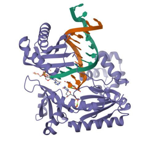
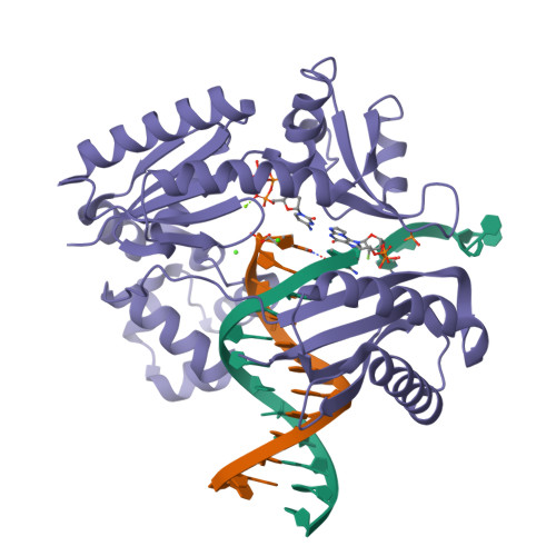
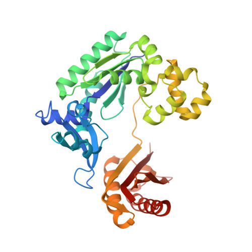
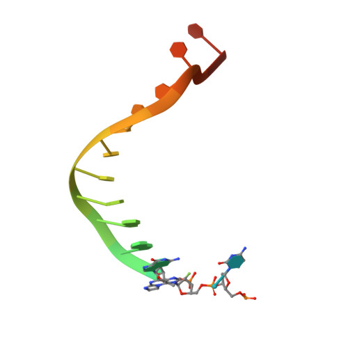
Replication of n(2) ,3-ethenoguanine by DNA polymerases.

Zhao, L.Christov, P.P.Kozekov, I.D.Pence, M.G.Pallan, P.S.Rizzo, C.J.Egli, M.Guengerich, F.P.

(2012) Angew Chem Int Ed Engl 51: 5466-5469


Macromolecules

Find similar proteins by:  (by identity cutoff)  |  3D Structure
Entity ID: 3
MoleculeChains Sequence LengthOrganismDetailsImage
DNA polymerase IVE [auth A],
F [auth B]
348Saccharolobus solfataricus P2Mutation(s): 0 
Gene Names: dbhdpo4SSO2448
EC: 2.7.7.7
UniProt
Find proteins for Q97W02 (Saccharolobus solfataricus (strain ATCC 35092 / DSM 1617 / JCM 11322 / P2))
Explore Q97W02 
Go to UniProtKB:  Q97W02
Entity Groups  
Sequence Clusters30% Identity50% Identity70% Identity90% Identity95% Identity100% Identity
UniProt GroupQ97W02
Sequence Annotations
Expand
  • Reference Sequence
Small Molecules
Ligands 3 Unique
IDChains Name / Formula / InChI Key2D Diagram3D Interactions
DCP
Query on DCP

Download Ideal Coordinates CCD File 
H [auth A],
L [auth B]
2'-DEOXYCYTIDINE-5'-TRIPHOSPHATE
C9 H16 N3 O13 P3
RGWHQCVHVJXOKC-SHYZEUOFSA-N
CA
Query on CA

Download Ideal Coordinates CCD File 
G [auth D],
K [auth A],
N [auth B]
CALCIUM ION
Ca
BHPQYMZQTOCNFJ-UHFFFAOYSA-N
MG
Query on MG

Download Ideal Coordinates CCD File 
I [auth A],
J [auth A],
M [auth B],
O [auth B]
MAGNESIUM ION
Mg
JLVVSXFLKOJNIY-UHFFFAOYSA-N
Experimental Data & Validation

Experimental Data

  • Method: X-RAY DIFFRACTION
  • Resolution: 2.30 Å
  • R-Value Free:  0.267 (Depositor), 0.280 (DCC) 
  • R-Value Work:  0.215 (Depositor), 0.230 (DCC) 
  • R-Value Observed: 0.218 (Depositor) 
Space Group: P 1 21 1
Unit Cell:
Length ( Å )Angle ( ˚ )
a = 52.532α = 90
b = 111.267β = 102.68
c = 98.942γ = 90
Software Package:
Software NamePurpose
HKL-2000data collection
REFMACrefinement
HKL-2000data reduction
HKL-2000data scaling
REFMACphasing

Structure Validation

View Full Validation Report


Ligand Structure Quality Assessment 

Created with Raphaël 2.3.0Worse 01 BetterLigand structure goodness of fit to experimental dataBest fitted DCPClick on this verticalbar to view details

Entry History 

Deposition Data

  • Released Date: 2012-04-25 
  • Deposition Author(s): Zhao, L.

Revision History  (Full details and data files)

  • Version 1.0: 2012-04-25
    Type: Initial release
  • Version 1.1: 2012-06-06
    Changes: Database references
  • Version 1.2: 2023-09-13
    Changes: Data collection, Database references, Derived calculations, Refinement description