6YAK | pdb_00006yak

Split gene transketolase, active alpha2beta2 heterotetramer


Experimental Data Snapshot

  • Method: X-RAY DIFFRACTION
  • Resolution: 1.34 Å
  • R-Value Free: 
    0.159 (Depositor), 0.160 (DCC) 
  • R-Value Work: 
    0.141 (Depositor), 0.140 (DCC) 

Starting Model: experimental
View more details

wwPDB Validation   3D Report Full Report


Ligand Structure Quality Assessment 

Created with Raphaël 2.3.0Worse 01 BetterLigand structure goodness of fit to experimental dataBest fitted 8ELClick on this verticalbar to view details

This is version 1.1 of the entry. See complete history


Literature

A 'Split-Gene' Transketolase From the Hyper-Thermophilic Bacterium Carboxydothermus hydrogenoformans : Structure and Biochemical Characterization.

James, P.Isupov, M.N.De Rose, S.A.Sayer, C.Cole, I.S.Littlechild, J.A.

(2020) Front Microbiol 11: 592353-592353

  • DOI: https://doi.org/10.3389/fmicb.2020.592353
  • Primary Citation of Related Structures:  
    6YAJ, 6YAK

  • PubMed Abstract: 

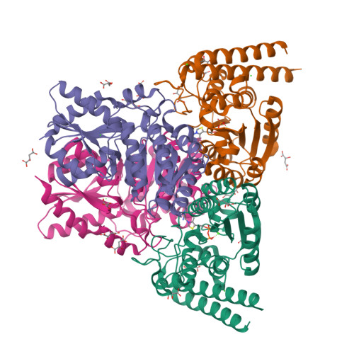
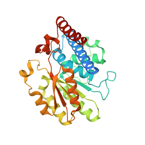
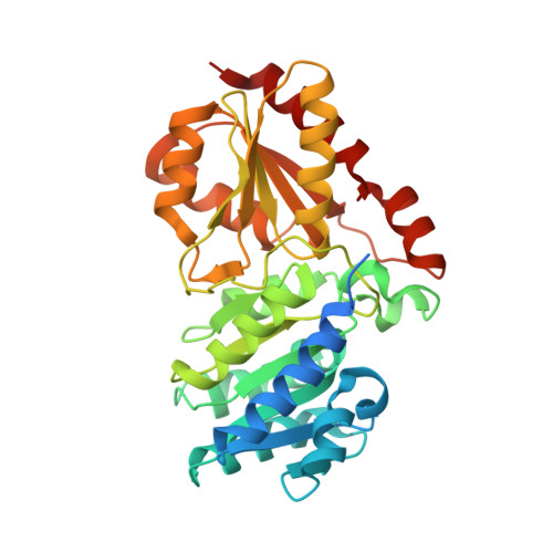
    A novel transketolase has been reconstituted from two separate polypeptide chains encoded by a 'split-gene' identified in the genome of the hyperthermophilic bacterium, Carboxydothermus hydrogenoformans . The reconstituted active α 2 β 2 tetrameric enzyme has been biochemically characterized and its activity has been determined using a range of aldehydes including glycolaldehyde, phenylacetaldehyde and cyclohexanecarboxaldehyde as the ketol acceptor and hydroxypyruvate as the donor. This reaction proceeds to near 100% completion due to the release of the product carbon dioxide and can be used for the synthesis of a range of sugars of interest to the pharmaceutical industry. This novel reconstituted transketolase is thermally stable with no loss of activity after incubation for 1 h at 70°C and is stable after 1 h incubation with 50% of the organic solvents methanol, ethanol, isopropanol, DMSO, acetonitrile and acetone. The X-ray structure of the holo reconstituted α 2 β 2 tetrameric transketolase has been determined to 1.4 Å resolution. In addition, the structure of an inactive tetrameric β 4 protein has been determined to 1.9 Å resolution. The structure of the active reconstituted α 2 β 2 enzyme has been compared to the structures of related enzymes; the E1 component of the pyruvate dehydrogenase complex and D-xylulose-5-phosphate synthase, in an attempt to rationalize differences in structure and substrate specificity between these enzymes. This is the first example of a reconstituted 'split-gene' transketolase to be biochemically and structurally characterized allowing its potential for industrial biocatalysis to be evaluated.


  • Organizational Affiliation

    Henry Wellcome Building for Biocatalysis, Biosciences, University of Exeter, Exeter, United Kingdom.


Macromolecules
Find similar proteins by:  (by identity cutoff)  |  3D Structure
Entity ID: 1
MoleculeChains Sequence LengthOrganismDetailsImage
N-terminal component of the split chain transketolaseA [auth AAA],
C [auth CCC]
309Carboxydothermus hydrogenoformansMutation(s): 0 
Entity Groups  
Sequence Clusters30% Identity50% Identity70% Identity90% Identity95% Identity100% Identity
Sequence Annotations
Expand
  • Reference Sequence
Find similar proteins by:  (by identity cutoff)  |  3D Structure
Entity ID: 2
MoleculeChains Sequence LengthOrganismDetailsImage
C-terminal component of the split chain transketolaseB [auth BBB],
D [auth DDD]
341Carboxydothermus hydrogenoformansMutation(s): 0 
Entity Groups  
Sequence Clusters30% Identity50% Identity70% Identity90% Identity95% Identity100% Identity
Sequence Annotations
Expand
  • Reference Sequence
Small Molecules
Ligands 5 Unique
IDChains Name / Formula / InChI Key2D Diagram3D Interactions
8EL (Subject of Investigation/LOI)
Query on 8EL

Download Ideal Coordinates CCD File 
E [auth AAA],
S [auth CCC]
2-[3-[(4-azanyl-2-methyl-pyrimidin-5-yl)methyl]-4-methyl-2H-1,3-thiazol-5-yl]ethyl phosphono hydrogen phosphate
C12 H20 N4 O7 P2 S
FCUXBJDJFWOWMD-UHFFFAOYSA-N
LMR
Query on LMR

Download Ideal Coordinates CCD File 
BA [auth DDD],
CA [auth DDD],
J [auth AAA],
K [auth AAA],
L [auth AAA]
(2S)-2-hydroxybutanedioic acid
C4 H6 O5
BJEPYKJPYRNKOW-REOHCLBHSA-N
MLT
Query on MLT

Download Ideal Coordinates CCD File 
H [auth AAA]
I [auth AAA]
N [auth BBB]
O [auth BBB]
P [auth BBB]
D-MALATE
C4 H6 O5
BJEPYKJPYRNKOW-UWTATZPHSA-N
CA (Subject of Investigation/LOI)
Query on CA

Download Ideal Coordinates CCD File 
F [auth AAA],
G [auth AAA],
T [auth CCC],
U [auth CCC]
CALCIUM ION
Ca
BHPQYMZQTOCNFJ-UHFFFAOYSA-N
CL
Query on CL

Download Ideal Coordinates CCD File 
AA [auth CCC],
DA [auth DDD],
M [auth AAA],
R [auth BBB]
CHLORIDE ION
Cl
VEXZGXHMUGYJMC-UHFFFAOYSA-M
Experimental Data & Validation

Experimental Data

  • Method: X-RAY DIFFRACTION
  • Resolution: 1.34 Å
  • R-Value Free:  0.159 (Depositor), 0.160 (DCC) 
  • R-Value Work:  0.141 (Depositor), 0.140 (DCC) 
Space Group: I 2 2 2
Unit Cell:
Length ( Å )Angle ( ˚ )
a = 123.078α = 90
b = 130.047β = 90
c = 165.897γ = 90
Software Package:
Software NamePurpose
REFMACrefinement
XDSdata reduction
XSCALEdata scaling
Aimlessdata scaling
MOLREPphasing
MoRDaphasing

Structure Validation

View Full Validation Report


Ligand Structure Quality Assessment 

Created with Raphaël 2.3.0Worse 01 BetterLigand structure goodness of fit to experimental dataBest fitted 8ELClick on this verticalbar to view details

Entry History & Funding Information

Deposition Data


Funding OrganizationLocationGrant Number
Biotechnology and Biological Sciences Research Council (BBSRC)United KingdomBB/L002035/1

Revision History  (Full details and data files)

  • Version 1.0: 2020-11-25
    Type: Initial release
  • Version 1.1: 2024-01-24
    Changes: Data collection, Database references, Derived calculations, Refinement description