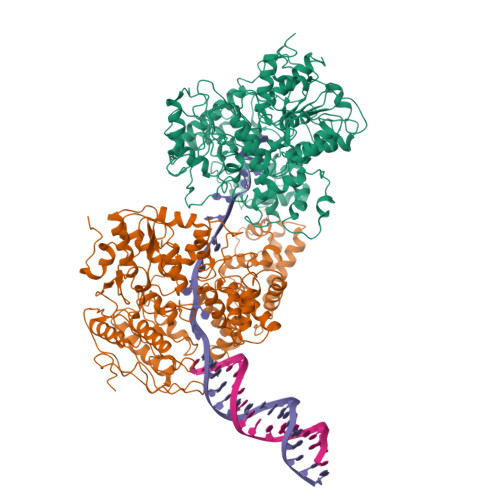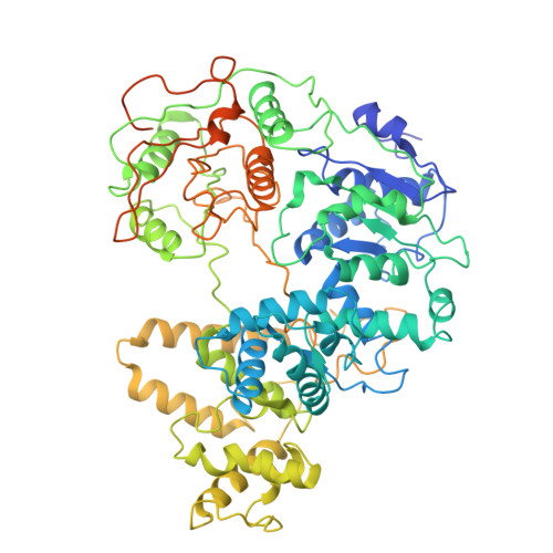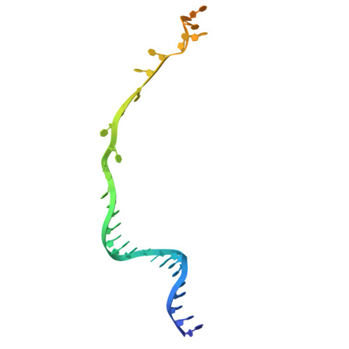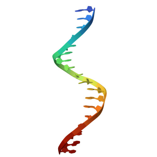
Structural basis for dimerization and activation of UvrD-family helicases.
Chadda, A., Nguyen, B., Lohman, T.M., Galburt, E.A.(2025) Proc Natl Acad Sci U S A 122: e2422330122-e2422330122
- PubMed: 40048277
- DOI: https://doi.org/10.1073/pnas.2422330122
- Primary Citation of Related Structures:
9DCI, 9DES - PubMed Abstract:
UvrD-family helicases are superfamily 1A motor proteins that function during DNA replication, recombination, repair, and transcription. UvrD family monomers translocate along single-stranded (ss) DNA but need to be activated by dimerization to unwind DNA in the absence of force or accessory factors. However, prior structural studies have only revealed monomeric complexes. Here, we report the first structures of a dimeric UvrD-family helicase, Mycobacterium tuberculosis UvrD1, both free and bound to a DNA junction. In each structure, the dimer interface occurs between the 2B subdomains of each subunit. The apo UvrD1 dimer is observed in symmetric compact and extended forms indicating substantial flexibility. This symmetry is broken in the DNA-bound dimer complex with leading and trailing subunits adopting distinct conformations. Biochemical experiments reveal that the Escherichia coli UvrD dimer shares the same 2B-2B interface. In contrast to the dimeric structures, an inactive, autoinhibited UvrD1 DNA-bound monomer structure reveals 2B subdomain-DNA contacts that are likely inhibitory. The major reorientation of the 2B subdomains that occurs upon UvrD1 dimerization prevents these duplex DNA interactions, thus relieving the autoinhibition. These structures reveal that the 2B subdomain serves a major regulatory role rather than participating directly in DNA unwinding.
Organizational Affiliation:
Department of Biochemistry and Molecular Biophysics, Washington University in Saint Louis School of Medicine, Saint Louis, MO 63110.