9EPP | pdb_00009epp

Cryo-EM Structure of Jumping Spider Rhodopsin-1 bound to a Giq heterotrimer


Experimental Data Snapshot

  • Method: ELECTRON MICROSCOPY
  • Resolution: 4.06 Å
  • Aggregation State: PARTICLE 
  • Reconstruction Method: SINGLE PARTICLE 

wwPDB Validation   3D Report Full Report


This is version 1.1 of the entry. See complete history


Literature

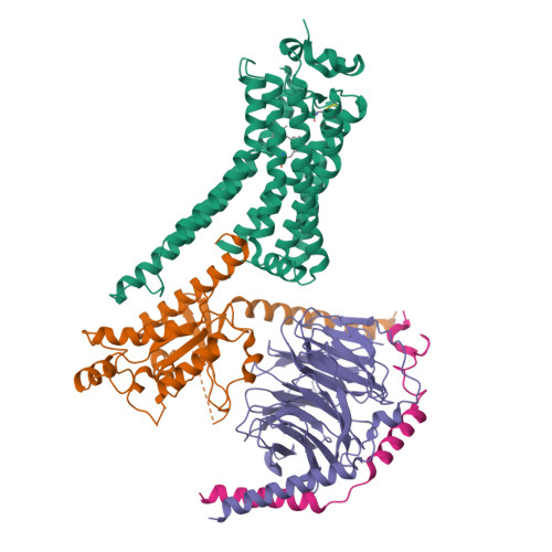
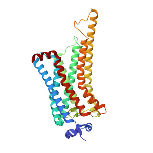
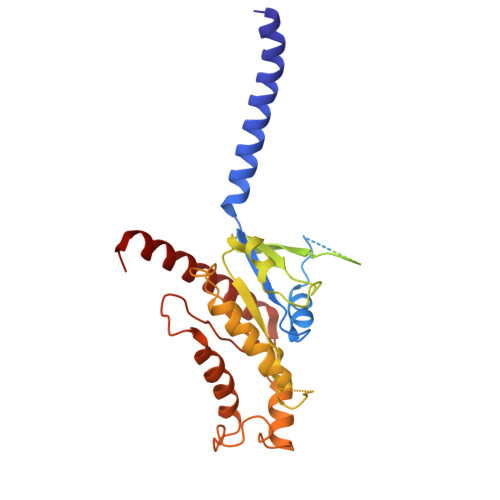
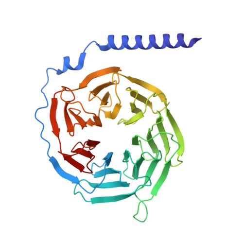
Active state structures of a bistable visual opsin bound to G proteins.

Tejero, O.Pamula, F.Koyanagi, M.Nagata, T.Afanasyev, P.Das, I.Deupi, X.Sheves, M.Terakita, A.Schertler, G.F.X.Rodrigues, M.J.Tsai, C.J.

(2024) Nat Commun 15: 8928-8928

  • DOI: https://doi.org/10.1038/s41467-024-53208-2
  • Primary Citation of Related Structures:  
    9EPP, 9EPQ, 9EPR

  • PubMed Abstract: 

    Opsins are G protein-coupled receptors (GPCRs) that have evolved to detect light stimuli and initiate intracellular signaling cascades. Their role as signal transducers is critical to light perception across the animal kingdom. Opsins covalently bind to the chromophore 11-cis retinal, which isomerizes to the all-trans isomer upon photon absorption, causing conformational changes that result in receptor activation. Monostable opsins, responsible for vision in vertebrates, release the chromophore after activation and must bind another retinal molecule to remain functional. In contrast, bistable opsins, responsible for non-visual light perception in vertebrates and for vision in invertebrates, absorb a second photon in the active state to return the chromophore and protein to the inactive state. Structures of bistable opsins in the activated state have proven elusive, limiting our understanding of how they function as bidirectional photoswitches. Here we present active state structures of a bistable opsin, jumping spider rhodopsin isoform-1 (JSR1), in complex with its downstream signaling partners, the G i and G q heterotrimers. These structures elucidate key differences in the activation mechanisms between monostable and bistable opsins, offering essential insights for the rational engineering of bistable opsins into diverse optogenetic tools to control G protein signaling pathways.


  • Organizational Affiliation

    Laboratory of Biomolecular Research, PSI Center for Life Sciences, Villigen-PSI, Switzerland.


Macromolecules
Find similar proteins by:  (by identity cutoff)  |  3D Structure
Entity ID: 1
MoleculeChains Sequence LengthOrganismDetailsImage
Kumopsin1A [auth R]327Hasarius adansoniMutation(s): 0 
Gene Names: HaRh1
UniProt
Find proteins for B1B1U5 (Hasarius adansoni)
Explore B1B1U5 
Go to UniProtKB:  B1B1U5
Entity Groups  
Sequence Clusters30% Identity50% Identity70% Identity90% Identity95% Identity100% Identity
UniProt GroupB1B1U5
Sequence Annotations
Expand
  • Reference Sequence
Find similar proteins by:  (by identity cutoff)  |  3D Structure
Entity ID: 2
MoleculeChains Sequence LengthOrganismDetailsImage
Guanine nucleotide-binding protein G(i) subunit alpha-1B [auth A]352Homo sapiensMutation(s): 11 
Gene Names: GNAI1
EC: 3.6.5
UniProt & NIH Common Fund Data Resources
Find proteins for P63096 (Homo sapiens)
Explore P63096 
Go to UniProtKB:  P63096
PHAROS:  P63096
GTEx:  ENSG00000127955 
Entity Groups  
Sequence Clusters30% Identity50% Identity70% Identity90% Identity95% Identity100% Identity
UniProt GroupP63096
Sequence Annotations
Expand
  • Reference Sequence
Find similar proteins by:  (by identity cutoff)  |  3D Structure
Entity ID: 3
MoleculeChains Sequence LengthOrganismDetailsImage
Guanine nucleotide-binding protein G(I)/G(S)/G(T) subunit beta-1C [auth B]339Homo sapiensMutation(s): 0 
Gene Names: GNB1
UniProt & NIH Common Fund Data Resources
Find proteins for P62873 (Homo sapiens)
Explore P62873 
Go to UniProtKB:  P62873
PHAROS:  P62873
GTEx:  ENSG00000078369 
Entity Groups  
Sequence Clusters30% Identity50% Identity70% Identity90% Identity95% Identity100% Identity
UniProt GroupP62873
Sequence Annotations
Expand
  • Reference Sequence
Find similar proteins by:  (by identity cutoff)  |  3D Structure
Entity ID: 4
MoleculeChains Sequence LengthOrganismDetailsImage
Guanine nucleotide-binding protein G(I)/G(S)/G(O) subunit gamma-2D [auth G]60Homo sapiensMutation(s): 0 
Gene Names: GNG2
UniProt & NIH Common Fund Data Resources
Find proteins for P59768 (Homo sapiens)
Explore P59768 
Go to UniProtKB:  P59768
PHAROS:  P59768
GTEx:  ENSG00000186469 
Entity Groups  
Sequence Clusters30% Identity50% Identity70% Identity90% Identity95% Identity100% Identity
UniProt GroupP59768
Sequence Annotations
Expand
  • Reference Sequence
Small Molecules
Ligands 1 Unique
IDChains Name / Formula / InChI Key2D Diagram3D Interactions
A1H6M (Subject of Investigation/LOI)
Query on A1H6M

Download Ideal Coordinates CCD File 
E [auth R]11,20-Ethanoretinal
C22 H30 O
FOYUFPJPFUUNOX-ZRTTZBFNSA-N
Experimental Data & Validation

Experimental Data

  • Method: ELECTRON MICROSCOPY
  • Resolution: 4.06 Å
  • Aggregation State: PARTICLE 
  • Reconstruction Method: SINGLE PARTICLE 

Structure Validation

View Full Validation Report


Entry History & Funding Information

Deposition Data


Funding OrganizationLocationGrant Number
European Research Council (ERC)European Union951644
H2020 Marie Curie Actions of the European CommissionEuropean Union701647
Swiss National Science FoundationSwitzerland192760
Swiss National Science FoundationSwitzerland18563

Revision History  (Full details and data files)

  • Version 1.0: 2024-10-23
    Type: Initial release
  • Version 1.1: 2024-11-06
    Changes: Data collection