Structural and biochemical characterization of recombinant wild type and a C30A mutant of trimethylamine dehydrogenase from methylophilus methylotrophus (sp. W(3)A(1)).
Trickey, P., Basran, J., Lian, L.Y., Chen, Z., Barton, J.D., Sutcliffe, M.J., Scrutton, N.S., Mathews, F.S.(2000) Biochemistry 39: 7678-7688
- PubMed: 10869173
- DOI: https://doi.org/10.1021/bi9927181
- Primary Citation of Related Structures:
1DJN, 1DJQ - PubMed Abstract:
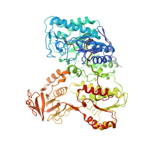
Trimethylamine dehydrogenase (TMADH) is an iron-sulfur flavoprotein that catalyzes the oxidative demethylation of trimethylamine to form dimethylamine and formaldehyde. It contains a unique flavin, in the form of a 6-S-cysteinyl FMN, which is bent by approximately 25 degrees along the N5-N10 axis of the flavin isoalloxazine ring. This unusual conformation is thought to modulate the properties of the flavin to facilitate catalysis, and has been postulated to be the result of covalent linkage to Cys-30 at the flavin C6 atom. We report here the crystal structures of recombinant wild-type and the C30A mutant TMADH enzymes, both determined at 2.2 A resolution. Combined crystallographic and NMR studies reveal the presence of inorganic phosphate in the FMN binding site in the deflavo fraction of both recombinant wild-type and C30A proteins. The presence of tightly bound inorganic phosphate in the recombinant enzymes explains the inability to reconstitute the deflavo forms of the recombinant wild-type and C30A enzymes that are generated in vivo. The active site structure and flavin conformation in C30A TMADH are identical to those in recombinant and native TMADH, thus revealing that, contrary to expectation, the 6-S-cysteinyl FMN link is not responsible for the 25 degrees butterfly bending along the N5-N10 axis of the flavin in TMADH. Computational quantum chemistry studies strongly support the proposed role of the butterfly bend in modulating the redox properties of the flavin. Solution studies reveal major differences in the kinetic behavior of the wild-type and C30A proteins. Computational studies reveal a hitherto, unrecognized, contribution made by the S(gamma) atom of Cys-30 to substrate binding, and a role for Cys-30 in the optimal geometrical alignment of substrate with the 6-S-cysteinyl FMN in the enzyme active site.
Organizational Affiliation:
Department of Biochemistry and Molecular Biophysics, Washington University School of Medicine, St. Louis, Missouri 63110, USA.


