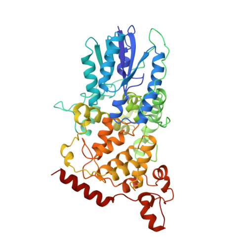
Crystal structure of DNA photolyase from Escherichia coli.
Park, H.W., Kim, S.T., Sancar, A., Deisenhofer, J.(1995) Science 268: 1866-1872
- PubMed: 7604260
- DOI: https://doi.org/10.1126/science.7604260
- Primary Citation of Related Structures:
1DNP - PubMed Abstract:
Photolyase repairs ultraviolet (UV) damage to DNA by splitting the cyclobutane ring of the major UV photoproduct, the cis, syn-cyclobutane pyrimidine dimer (Pyr <> Pyr). The reaction is initiated by blue light and proceeds through long-range energy transfer, single electron transfer, and enzyme catalysis by a radical mechanism. The three-dimensional crystallographic structure of DNA photolyase from Escherichia coli is presented and the atomic model was refined to an R value of 0.172 at 2.3 A resolution. The polypeptide chain of 471 amino acids is folded into an amino-terminal alpha/beta domain resembling dinucleotide binding domains and a carboxyl-terminal helical domain; a loop of 72 residues connects the domains. The light-harvesting cofactor 5,10-methenyltetrahydrofolylpolyglutamate (MTHF) binds in a cleft between the two domains. Energy transfer from MTHF to the catalytic cofactor flavin adenine dinucleotide (FAD) occurs over a distance of 16.8 A. The FAD adopts a U-shaped conformation between two helix clusters in the center of the helical domain and is accessible through a hole in the surface of this domain. Dimensions and polarity of the hole match those of a Pyr <> Pyr dinucleotide, suggesting that the Pyr <> Pyr "flips out" of the helix to fit into this hole, and that electron transfer between the flavin and the Pyr <> Pyr occurs over van der Waals contact distance.
- Department of Biochemistry, University of Texas Southwestern Medical Center, Dallas 75235, USA.
Organizational Affiliation:


