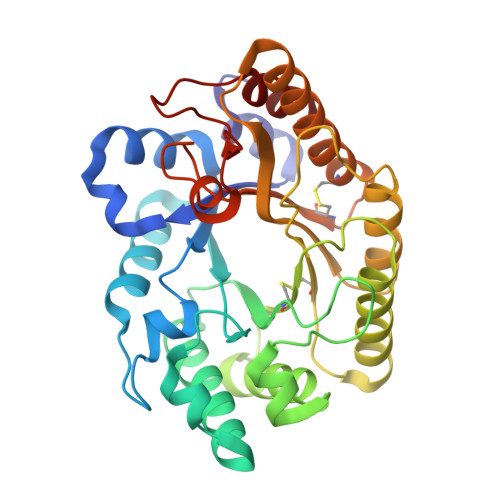
1FHD

CRYSTAL STRUCTURE OF THE XYLANASE CEX WITH XYLOBIOSE-DERIVED IMIDAZOLE INHIBITOR


Experimental Data Snapshot

  • Method: X-RAY DIFFRACTION
  • Resolution: 1.90 Å
  • R-Value Free: 0.219 
  • R-Value Work: 0.193 
  • R-Value Observed: 0.193 

wwPDB Validation   3D Report Full Report


Ligand Structure Quality Assessment 


This is version 2.1 of the entry. See complete history


Literature

Detailed structural analysis of glycosidase/inhibitor interactions: complexes of Cex from Cellulomonas fimi with xylobiose-derived aza-sugars.

Notenboom, V.Williams, S.J.Hoos, R.Withers, S.G.Rose, D.R.

(2000) Biochemistry 39: 11553-11563

  • DOI: https://doi.org/10.1021/bi0010625
  • Primary Citation of Related Structures:  
    1FH7, 1FH8, 1FH9, 1FHD

  • PubMed Abstract: 

    Detailed insights into the mode of binding of a series of tight-binding aza-sugar glycosidase inhibitors of two fundamentally different classes are described through X-ray crystallographic studies of complexes with the retaining family 10 xylanase Cex from Cellulomonas fimi. Complexes with xylobiose-derived aza-sugar inhibitors of the substituted "amidine" class (xylobio-imidazole, K(i) = 150 nM; xylobio-lactam oxime, K(i) = 370 nM) reveal lateral interaction of the "glycosidic" nitrogen with the acid/base catalyst (Glu127) and hydrogen bonding of the sugar 2-hydroxyl with the catalytic nucleophile (Glu233), as expected. Tight binding of xylobio-isofagomine (K(i) = 130 nM) appears to be a consequence of strong interactions of the ring nitrogen with the catalytic nucleophile while, surprisingly, no direct protein contacts are made with the ring nitrogen of the xylobio-deoxynojirimycin analogue (K(i) = 5800 nM). Instead the nitrogen interacts with two ordered water molecules, thereby accounting for its relatively weaker binding, though it still binds some 1200-fold more tightly than does xylobiose, presumably as a consequence of electrostatic interactions at the active site. Dramatically weaker binding of these same inhibitors to the family 11 xylanase Bcx from Bacillus circulans (K(i) from 0.5 to 1.5 mM) is rationalized for the substituted amidines on the basis that this enzyme utilizes a syn protonation trajectory and likely hydrolyzes via a (2,5)B boat transition state. Weaker binding of the deoxynojirimycin and isofagomine analogues likely reflects the energetic penalty for distortion of these analogues to a (2,5)B conformation, possibly coupled with destabilizing interactions with Tyr69, a conserved, catalytically essential active site residue.


  • Organizational Affiliation

    Protein Engineering Network of Centres of Excellence, Ontario Cancer Institute and Department of Medical Biophysics, University of Toronto, 610 University Avenue, Toronto, Ontario, Canada.


Macromolecules
Find similar proteins by:  (by identity cutoff)  |  3D Structure
Entity ID: 1
MoleculeChains Sequence LengthOrganismDetailsImage
BETA-1,4-XYLANASE312Cellulomonas fimiMutation(s): 0 
EC: 3.2.1.91 (PDB Primary Data), 3.2.1.8 (UniProt)
UniProt
Find proteins for P07986 (Cellulomonas fimi)
Explore P07986 
Go to UniProtKB:  P07986
Entity Groups  
Sequence Clusters30% Identity50% Identity70% Identity90% Identity95% Identity100% Identity
UniProt GroupP07986
Sequence Annotations
Expand
  • Reference Sequence
Small Molecules
Ligands 2 Unique
IDChains Name / Formula / InChI Key2D Diagram3D Interactions
XIM
Query on XIM

Download Ideal Coordinates CCD File 
C [auth A]5,6,7,8-TETRAHYDRO-IMIDAZO[1,2-A]PYRIDINE-6,7,8-TRIOL
C7 H10 N2 O3
QMHSBFMMOLGPGR-NGJCXOISSA-N
XYP
Query on XYP

Download Ideal Coordinates CCD File 
B [auth A]beta-D-xylopyranose
C5 H10 O5
SRBFZHDQGSBBOR-KKQCNMDGSA-N
Experimental Data & Validation

Experimental Data

  • Method: X-RAY DIFFRACTION
  • Resolution: 1.90 Å
  • R-Value Free: 0.219 
  • R-Value Work: 0.193 
  • R-Value Observed: 0.193 
  • Space Group: P 41 21 2
Unit Cell:
Length ( Å )Angle ( ˚ )
a = 85.343α = 90
b = 85.343β = 90
c = 78.604γ = 90
Software Package:
Software NamePurpose
MAR345data collection
SCALEPACKdata scaling
CNSrefinement
CNSphasing

Structure Validation

View Full Validation Report


Ligand Structure Quality Assessment 


Entry History 

Deposition Data

Revision History  (Full details and data files)

  • Version 1.0: 2000-08-23
    Type: Initial release
  • Version 1.1: 2008-04-27
    Changes: Version format compliance
  • Version 1.2: 2011-07-13
    Changes: Version format compliance
  • Version 1.3: 2017-10-04
    Changes: Refinement description
  • Version 2.0: 2020-07-29
    Type: Remediation
    Reason: Carbohydrate remediation
    Changes: Atomic model, Data collection, Derived calculations, Structure summary
  • Version 2.1: 2024-10-16
    Changes: Data collection, Database references, Derived calculations, Structure summary