1FT1

CRYSTAL STRUCTURE OF PROTEIN FARNESYLTRANSFERASE AT 2.25 ANGSTROMS RESOLUTION


Experimental Data Snapshot

  • Method: X-RAY DIFFRACTION
  • Resolution: 2.25 Å
  • R-Value Free: 0.260 
  • R-Value Work: 0.210 
  • R-Value Observed: 0.210 

wwPDB Validation   3D Report Full Report


This is version 1.3 of the entry. See complete history


Literature

Crystal structure of protein farnesyltransferase at 2.25 angstrom resolution.

Park, H.W.Boduluri, S.R.Moomaw, J.F.Casey, P.J.Beese, L.S.

(1997) Science 275: 1800-1804

  • DOI: https://doi.org/10.1126/science.275.5307.1800
  • Primary Citation of Related Structures:  
    1FT1

  • PubMed Abstract: 

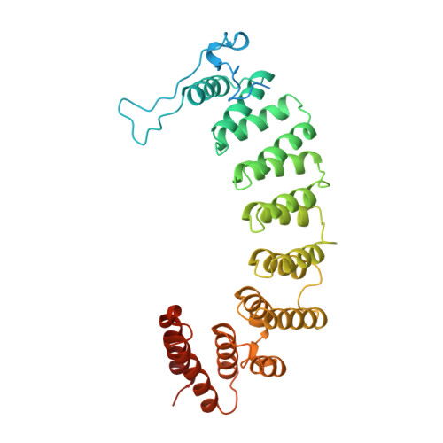
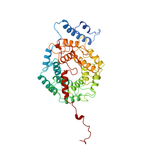
    Protein farnesyltransferase (FTase) catalyzes the carboxyl-terminal lipidation of Ras and several other cellular signal transduction proteins. The essential nature of this modification for proper function of these proteins has led to the emergence of FTase as a target for the development of new anticancer therapy. Inhibition of this enzyme suppresses the transformed phenotype in cultured cells and causes tumor regression in animal models. The crystal structure of heterodimeric mammalian FTase was determined at 2.25 angstrom resolution. The structure shows a combination of two unusual domains: a crescent-shaped seven-helical hairpin domain and an alpha-alpha barrel domain. The active site is formed by two clefts that intersect at a bound zinc ion. One cleft contains a nine-residue peptide that may mimic the binding of the Ras substrate; the other cleft is lined with highly conserved aromatic residues appropriate for binding the farnesyl isoprenoid with required specificity.


  • Organizational Affiliation

    Department of Biochemistry, Duke University Medical Center, Durham, NC 27710, USA.


Macromolecules
Find similar proteins by:  (by identity cutoff)  |  3D Structure
Entity ID: 1
MoleculeChains Sequence LengthOrganismDetailsImage
PROTEIN FARNESYLTRANSFERASE377Rattus norvegicusMutation(s): 0 
Gene Names: CDNA
EC: 2.5.1 (PDB Primary Data), 2.5.1.58 (UniProt), 2.5.1.59 (UniProt)
UniProt
Find proteins for Q04631 (Rattus norvegicus)
Explore Q04631 
Go to UniProtKB:  Q04631
Entity Groups  
Sequence Clusters30% Identity50% Identity70% Identity90% Identity95% Identity100% Identity
UniProt GroupQ04631
Sequence Annotations
Expand
  • Reference Sequence
Find similar proteins by:  (by identity cutoff)  |  3D Structure
Entity ID: 2
MoleculeChains Sequence LengthOrganismDetailsImage
PROTEIN FARNESYLTRANSFERASE437Rattus norvegicusMutation(s): 0 
Gene Names: CDNA
EC: 2.5.1 (PDB Primary Data), 2.5.1.58 (UniProt)
UniProt
Find proteins for Q02293 (Rattus norvegicus)
Explore Q02293 
Go to UniProtKB:  Q02293
Entity Groups  
Sequence Clusters30% Identity50% Identity70% Identity90% Identity95% Identity100% Identity
UniProt GroupQ02293
Sequence Annotations
Expand
  • Reference Sequence
Small Molecules
Ligands 1 Unique
IDChains Name / Formula / InChI Key2D Diagram3D Interactions
ZN
Query on ZN

Download Ideal Coordinates CCD File 
C [auth B]ZINC ION
Zn
PTFCDOFLOPIGGS-UHFFFAOYSA-N
Experimental Data & Validation

Experimental Data

  • Method: X-RAY DIFFRACTION
  • Resolution: 2.25 Å
  • R-Value Free: 0.260 
  • R-Value Work: 0.210 
  • R-Value Observed: 0.210 
  • Space Group: P 65
Unit Cell:
Length ( Å )Angle ( ˚ )
a = 167.06α = 90
b = 167.06β = 90
c = 97.9γ = 120
Software Package:
Software NamePurpose
X-PLORmodel building
X-PLORrefinement
DENZOdata reduction
SCALEPACKdata scaling
X-PLORphasing

Structure Validation

View Full Validation Report


Entry History 

Deposition Data

Revision History  (Full details and data files)

  • Version 1.0: 1998-03-18
    Type: Initial release
  • Version 1.1: 2008-03-24
    Changes: Version format compliance
  • Version 1.2: 2011-07-13
    Changes: Version format compliance
  • Version 1.3: 2024-02-07
    Changes: Data collection, Database references, Derived calculations, Other