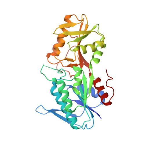
1IBS

PHOSPHORIBOSYLDIPHOSPHATE SYNTHETASE IN COMPLEX WITH CADMIUM IONS


Experimental Data Snapshot

  • Method: X-RAY DIFFRACTION
  • Resolution: 2.80 Å
  • R-Value Free: 0.273 
  • R-Value Work: 0.200 
  • R-Value Observed: 0.200 

wwPDB Validation   3D Report Full Report


Ligand Structure Quality Assessment 


This is version 1.4 of the entry. See complete history


Literature

Binding of cations in Bacillus subtilis phosphoribosyldiphosphate synthetase and their role in catalysis.

Eriksen, T.A.Kadziola, A.Larsen, S.

(2002) Protein Sci 11: 271-279

  • DOI: https://doi.org/10.1110/ps.28502
  • Primary Citation of Related Structures:  
    1IBS

  • PubMed Abstract: 

    The binding sites for the two cations essential for the catalytic function of 5-phospho-D-ribosyl alpha-1-diphosphate (PRPP) synthases have been identified from the structure of the Bacillus subtilis phosphoribosyldiphosphate synthetase (PRPPsase) with bound Cd(2+). The structure determined from X-ray diffraction data to 2.8-A resolution reveals the same hexameric arrangement of the subunits that was observed in the complexes of the enzyme with the activator sulfate and the allosteric inhibitor ADP. Two cation binding sites were localized in each of the two domains of the subunits that compose the hexamer; each domain of the subunit has an associated cation. In addition to the bound Cd(2+), the Cd(2+)-PRPPsase structure contains a sulfate ion in the regulatory site, a sulfate ion at the ribose-5-phosphate binding site, and an AMP moiety at the ATP binding site. Comparison of the Cd(2+)-PRPPsase to the structures of the PRPPsase complexed with sulfate and mADP reveals the structural rearrangement induced by the binding of the free cation, which is essential for the initiation of the reaction. The comparison to the cPRPP complex of glutamine phosphoribosylpyrophosphate amidotransferase from Escherichia coli, a type I phosphoribosyltransferase, provided information about the binding of PRPP. This strongly indicates that the binding of both substrates must lead to a stabilized conformation of the loop region, which remains unresolved in the known PRPPsase complex structures.


  • Organizational Affiliation

    Centre for Crystallographic Studies, Department of Chemistry, University of Copenhagen, 2100 Copenhagen, Denmark.


Macromolecules
Find similar proteins by:  (by identity cutoff)  |  3D Structure
Entity ID: 1
MoleculeChains Sequence LengthOrganismDetailsImage
RIBOSE-PHOSPHATE PYROPHOSPHOKINASE
A, B
317Bacillus subtilisMutation(s): 0 
Gene Names: PRS
EC: 2.7.6.1
UniProt
Find proteins for P14193 (Bacillus subtilis (strain 168))
Explore P14193 
Go to UniProtKB:  P14193
Entity Groups  
Sequence Clusters30% Identity50% Identity70% Identity90% Identity95% Identity100% Identity
UniProt GroupP14193
Sequence Annotations
Expand
  • Reference Sequence
Small Molecules
Ligands 3 Unique
IDChains Name / Formula / InChI Key2D Diagram3D Interactions
ABM
Query on ABM

Download Ideal Coordinates CCD File 
G [auth A],
L [auth B]
METHYL PHOSPHONIC ACID ADENOSINE ESTER
C11 H16 N5 O6 P
PXSSQXBLDTZHLF-IOSLPCCCSA-N
CD
Query on CD

Download Ideal Coordinates CCD File 
E [auth A],
F [auth A],
J [auth B],
K [auth B]
CADMIUM ION
Cd
WLZRMCYVCSSEQC-UHFFFAOYSA-N
SO4
Query on SO4

Download Ideal Coordinates CCD File 
C [auth A],
D [auth A],
H [auth B],
I [auth B]
SULFATE ION
O4 S
QAOWNCQODCNURD-UHFFFAOYSA-L
Experimental Data & Validation

Experimental Data

  • Method: X-RAY DIFFRACTION
  • Resolution: 2.80 Å
  • R-Value Free: 0.273 
  • R-Value Work: 0.200 
  • R-Value Observed: 0.200 
  • Space Group: P 63
Unit Cell:
Length ( Å )Angle ( ˚ )
a = 115.6α = 90
b = 115.6β = 90
c = 107.67γ = 120
Software Package:
Software NamePurpose
X-PLORmodel building
X-PLORrefinement
CCP4data scaling
ROTAVATAdata scaling
TRUNCATEdata scaling
X-PLORphasing

Structure Validation

View Full Validation Report


Ligand Structure Quality Assessment 


Entry History 

Deposition Data

Revision History  (Full details and data files)

  • Version 1.0: 2002-02-06
    Type: Initial release
  • Version 1.1: 2008-04-27
    Changes: Version format compliance
  • Version 1.2: 2011-07-13
    Changes: Derived calculations, Version format compliance
  • Version 1.3: 2017-10-04
    Changes: Refinement description
  • Version 1.4: 2024-02-07
    Changes: Data collection, Database references, Derived calculations