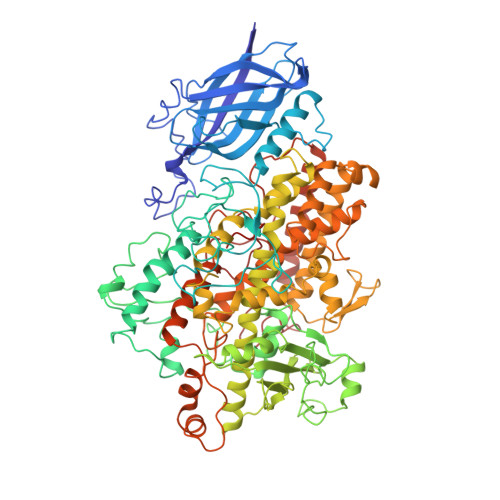
1JNQ

LIPOXYGENASE-3 (SOYBEAN) COMPLEX WITH EPIGALLOCATHECHIN (EGC)


Experimental Data Snapshot

  • Method: X-RAY DIFFRACTION
  • Resolution: 2.10 Å
  • R-Value Free: 0.295 
  • R-Value Work: 0.199 
  • R-Value Observed: 0.232 

Starting Model: experimental
View more details

wwPDB Validation   3D Report Full Report


Ligand Structure Quality Assessment 


This is version 1.5 of the entry. See complete history


Literature

Inhibition of lipoxygenase by (-)-epigallocatechin gallate: X-ray analysis at 2.1 A reveals degradation of EGCG and shows soybean LOX-3 complex with EGC instead.

Skrzypczak-Jankun, E.Zhou, K.Jankun, J.

(2003) Int J Mol Med 12: 415-420

  • Primary Citation of Related Structures:  
    1JNQ

  • PubMed Abstract: 

    Lipoxygenases (LOXs) are non-heme iron containing enzymes ubiquitous in nature and participating in the metabolism of the polyunsaturated fatty acids (PUFA). They are capable of combining their dioxygenase activity with its co-oxidative activity manifesting itself in biotransformation reactions catalyzed by LOXs for other than PUFA small molecules. LOXs involvement in inflammatory diseases and cancer have been well documented. Catechins are the natural flavonoids of known inhibitory activity toward dioxygenases with a potential to be utilized in disease prevention and treatment. This work presents results obtained from an X-ray analysis of (-)-epigallocatechin gallate (EGCG) interacting with soybean lipoxygenase-3. The 3D structure of the resulting complex reveals the inhibitor depicting (-)-epigallo-catechin that lacks galloyl moiety. The A-ring is near the iron co-factor, attached by the hydrogen bond to the C-terminus of the enzyme, and the B-ring hydroxyl groups participate in the hydrogen bonds and the van der Waals interactions formed by the surrounding amino acids and water molecules.


  • Organizational Affiliation

    Urology Research Center, Medical College of Ohio, Toledo, OH 43614-5807, USA. eskrzypczak@mco.edu


Macromolecules
Find similar proteins by:  (by identity cutoff)  |  3D Structure
Entity ID: 1
MoleculeChains Sequence LengthOrganismDetailsImage
lipoxygenase-3857Glycine maxMutation(s): 0 
EC: 1.13.11.12 (PDB Primary Data), 1.13.11.58 (UniProt)
UniProt
Find proteins for P09186 (Glycine max)
Explore P09186 
Go to UniProtKB:  P09186
Entity Groups  
Sequence Clusters30% Identity50% Identity70% Identity90% Identity95% Identity100% Identity
UniProt GroupP09186
Sequence Annotations
Expand
  • Reference Sequence
Small Molecules
Ligands 2 Unique
IDChains Name / Formula / InChI Key2D Diagram3D Interactions
EGT
Query on EGT

Download Ideal Coordinates CCD File 
C [auth A]2-(3,4,5-TRIHYDROXY-PHENYL)-CHROMAN-3,5,7-TRIOL
C15 H14 O7
XMOCLSLCDHWDHP-IUODEOHRSA-N
FE2
Query on FE2

Download Ideal Coordinates CCD File 
B [auth A]FE (II) ION
Fe
CWYNVVGOOAEACU-UHFFFAOYSA-N
Experimental Data & Validation

Experimental Data

  • Method: X-RAY DIFFRACTION
  • Resolution: 2.10 Å
  • R-Value Free: 0.295 
  • R-Value Work: 0.199 
  • R-Value Observed: 0.232 
  • Space Group: C 1 2 1
Unit Cell:
Length ( Å )Angle ( ˚ )
a = 112.73α = 90
b = 137.27β = 95.53
c = 61.88γ = 90
Software Package:
Software NamePurpose
DENZOdata reduction
SCALEPACKdata scaling
X-PLORmodel building
X-PLORrefinement
X-PLORphasing

Structure Validation

View Full Validation Report


Ligand Structure Quality Assessment 


Entry History 

Deposition Data

Revision History  (Full details and data files)

  • Version 1.0: 2003-06-03
    Type: Initial release
  • Version 1.1: 2007-10-16
    Changes: Version format compliance
  • Version 1.2: 2011-07-13
    Changes: Version format compliance
  • Version 1.3: 2018-01-31
    Changes: Database references
  • Version 1.4: 2018-04-04
    Changes: Data collection
  • Version 1.5: 2023-08-16
    Changes: Data collection, Database references, Derived calculations, Refinement description