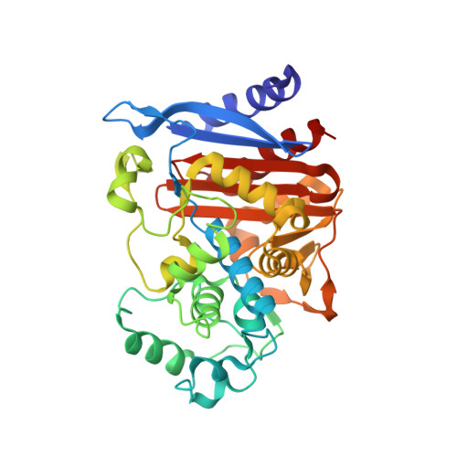
1LLB

Crystal Structure Of AmpC beta-Lactamase From E. Coli In Complex With ATMO-penicillin


Experimental Data Snapshot

  • Method: X-RAY DIFFRACTION
  • Resolution: 1.72 Å
  • R-Value Free: 0.199 
  • R-Value Work: 0.178 

Starting Model: experimental
View more details

wwPDB Validation   3D Report Full Report


Ligand Structure Quality Assessment 


This is version 1.4 of the entry. See complete history


Literature

Using steric hindrance to design new inhibitors of class C beta-lactamases.

Trehan, I.Morandi, F.Blaszczak, L.C.Shoichet, B.K.

(2002) Chem Biol 9: 971-980

  • DOI: https://doi.org/10.1016/s1074-5521(02)00211-9
  • Primary Citation of Related Structures:  
    1LL9, 1LLB

  • PubMed Abstract: 

    beta-lactamases confer resistance to beta-lactam antibiotics such as penicillins and cephalosporins. However, beta-lactams that form an acyl-intermediate with the enzyme but subsequently are hindered from forming a catalytically competent conformation seem to be inhibitors of beta-lactamases. This inhibition may be imparted by specific groups on the ubiquitous R(1) side chain of beta-lactams, such as the 2-amino-4-thiazolyl methoxyimino (ATMO) group common among third-generation cephalosporins. Using steric hindrance of deacylation as a design guide, penicillin and carbacephem substrates were converted into effective beta-lactamase inhibitors and antiresistance antibiotics. To investigate the structural bases of inhibition, the crystal structures of the acyl-adducts of the penicillin substrate amoxicillin and the new analogous inhibitor ATMO-penicillin were determined. ATMO-penicillin binds in a catalytically incompetent conformation resembling that adopted by third-generation cephalosporins, demonstrating the transferability of such sterically hindered groups in inhibitor design.


  • Organizational Affiliation

    Department of Molecular Pharmacology and Biological Chemistry, Northwestern University, 303 E Chicago Avenue, Chicago, IL 60611, USA.


Macromolecules
Find similar proteins by:  (by identity cutoff)  |  3D Structure
Entity ID: 1
MoleculeChains Sequence LengthOrganismDetailsImage
beta-lactamase
A, B
358Escherichia coliMutation(s): 0 
Gene Names: ampC
EC: 3.5.2.6
UniProt
Find proteins for P00811 (Escherichia coli (strain K12))
Explore P00811 
Go to UniProtKB:  P00811
Entity Groups  
Sequence Clusters30% Identity50% Identity70% Identity90% Identity95% Identity100% Identity
UniProt GroupP00811
Sequence Annotations
Expand
  • Reference Sequence
Small Molecules
Ligands 1 Unique
IDChains Name / Formula / InChI Key2D Diagram3D Interactions
PCN
Query on PCN

Download Ideal Coordinates CCD File 
C [auth B]2-{1-[2-(2-AMINO-THIAZOL-4-YL)-2-METHOXYIMINO-ACETYLAMINO]-2-OXO-ETHYL}-5,5-DIMETHYL-THIAZOLIDINE-4-CARBOXYLIC ACID
C14 H19 N5 O5 S2
YVNKGXXVZIQNIV-RKMXGKDGSA-N
Binding Affinity Annotations 
IDSourceBinding Affinity
PCN PDBBind:  1LLB IC50: 900 (nM) from 1 assay(s)
Experimental Data & Validation

Experimental Data

  • Method: X-RAY DIFFRACTION
  • Resolution: 1.72 Å
  • R-Value Free: 0.199 
  • R-Value Work: 0.178 
  • Space Group: C 1 2 1
Unit Cell:
Length ( Å )Angle ( ˚ )
a = 118.663α = 90
b = 76.734β = 116.186
c = 98.169γ = 90
Software Package:
Software NamePurpose
DENZOdata reduction
SCALEPACKdata scaling
CNSrefinement
CNSphasing

Structure Validation

View Full Validation Report


Ligand Structure Quality Assessment 


Entry History 

Deposition Data

Revision History  (Full details and data files)

  • Version 1.0: 2002-10-02
    Type: Initial release
  • Version 1.1: 2008-04-28
    Changes: Version format compliance
  • Version 1.2: 2011-07-13
    Changes: Version format compliance
  • Version 1.3: 2023-08-16
    Changes: Data collection, Database references, Derived calculations, Refinement description
  • Version 1.4: 2024-11-06
    Changes: Structure summary