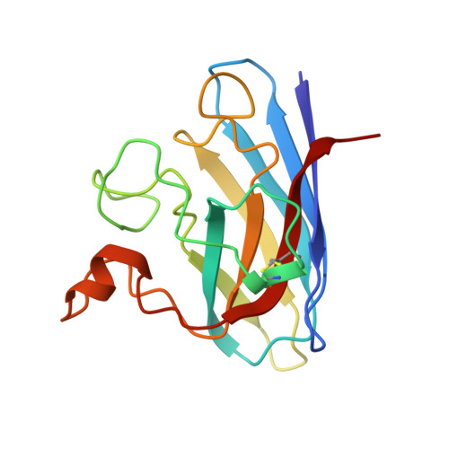
1MFM

MONOMERIC HUMAN SOD MUTANT F50E/G51E/E133Q AT ATOMIC RESOLUTION


Experimental Data Snapshot

  • Method: X-RAY DIFFRACTION
  • Resolution: 1.02 Å
  • R-Value Observed: 0.118 

Starting Model: experimental
View more details

wwPDB Validation   3D Report Full Report


This is version 1.5 of the entry. See complete history


Literature

The crystal structure of the monomeric human SOD mutant F50E/G51E/E133Q at atomic resolution. The enzyme mechanism revisited.

Ferraroni, M.Rypniewski, W.Wilson, K.S.Viezzoli, M.S.Banci, L.Bertini, I.Mangani, S.

(1999) J Mol Biol 288: 413-426

  • DOI: https://doi.org/10.1006/jmbi.1999.2681
  • Primary Citation of Related Structures:  
    1MFM

  • PubMed Abstract: 

    The crystal structure of the engineered monomeric human Cu,ZnSOD triple mutant F50E/G51E/E133Q (Q133M2SOD) is reported at atomic resolution (1.02 A). This derivative has about 20 % of the wild-type activity. Crystals of Q133M2SOD have been obtained in the presence of CdCl2. The metal binding site is disordered, with both cadmium and copper ions simultaneously binding to the copper site. The cadmium (II) ions occupy about 45 % of the copper sites by binding the four histidine residues which ligate copper in the native enzyme, and two further water molecules to complete octahedral coordination. The copper ion is tri-coordinate, and the fourth histidine (His63) is detached from copper and bridges cadmium and zinc. X-ray absorption spectroscopy performed on the crystals suggests that the copper ion has undergone partial photoreduction upon exposure to the synchrotron light. The structure is also disordered in the disulfide bridge region of loop IV that is located at the subunit/subunit interface in the native SOD dimer. As a consequence, the catalytically relevant Arg143 residue is disordered. The present structure has been compared to other X-ray structures on various isoenzymes and to the solution structure of the same monomeric form. The structural results suggest that the low activity of monomeric SOD is due to the disorder in the conformation of the side-chain of Arg143 as well as of loop IV. It is proposed that the subunit-subunit interactions in the multimeric forms of the enzyme are needed to stabilize the correct geometry of the cavity and the optimal orientation of the charged residues in the active channel. Furthermore, the different coordination of cadmium and copper ions, contemporaneously present in the same site, are taken as models for the oxidized and reduced copper species, respectively. These properties of the structure have allowed us to revisit the enzymatic mechanism.


  • Organizational Affiliation

    Department of Chemistry, University of Florence, Via Gino Capponi 9, Florence, I-50121, Italy.


Macromolecules
Find similar proteins by:  (by identity cutoff)  |  3D Structure
Entity ID: 1
MoleculeChains Sequence LengthOrganismDetailsImage
PROTEIN (COPPER,ZINC SUPEROXIDE DISMUTASE)153Homo sapiensMutation(s): 5 
Gene Names: SOD1
EC: 1.15.1.1
UniProt & NIH Common Fund Data Resources
Find proteins for P00441 (Homo sapiens)
Explore P00441 
Go to UniProtKB:  P00441
PHAROS:  P00441
GTEx:  ENSG00000142168 
Entity Groups  
Sequence Clusters30% Identity50% Identity70% Identity90% Identity95% Identity100% Identity
UniProt GroupP00441
Sequence Annotations
Expand
  • Reference Sequence
Small Molecules
Ligands 4 Unique
IDChains Name / Formula / InChI Key2D Diagram3D Interactions
CD
Query on CD

Download Ideal Coordinates CCD File 
D [auth A]
E [auth A]
F [auth A]
G [auth A]
H [auth A]
D [auth A],
E [auth A],
F [auth A],
G [auth A],
H [auth A],
I [auth A],
J [auth A],
K [auth A],
L [auth A]
CADMIUM ION
Cd
WLZRMCYVCSSEQC-UHFFFAOYSA-N
ZN
Query on ZN

Download Ideal Coordinates CCD File 
B [auth A]ZINC ION
Zn
PTFCDOFLOPIGGS-UHFFFAOYSA-N
CU
Query on CU

Download Ideal Coordinates CCD File 
C [auth A]COPPER (II) ION
Cu
JPVYNHNXODAKFH-UHFFFAOYSA-N
CL
Query on CL

Download Ideal Coordinates CCD File 
M [auth A],
N [auth A]
CHLORIDE ION
Cl
VEXZGXHMUGYJMC-UHFFFAOYSA-M
Experimental Data & Validation

Experimental Data

  • Method: X-RAY DIFFRACTION
  • Resolution: 1.02 Å
  • R-Value Observed: 0.118 
  • Space Group: P 21 21 21
Unit Cell:
Length ( Å )Angle ( ˚ )
a = 34.99α = 90
b = 48.11β = 90
c = 81.08γ = 90
Software Package:
Software NamePurpose
DENZOdata reduction
SCALEPACKdata scaling
AMoREphasing
SHELXLrefinement

Structure Validation

View Full Validation Report


Entry History 

Deposition Data

Revision History  (Full details and data files)

  • Version 1.0: 1999-04-21
    Type: Initial release
  • Version 1.1: 2007-10-16
    Changes: Version format compliance
  • Version 1.2: 2011-07-13
    Changes: Version format compliance
  • Version 1.3: 2017-10-04
    Changes: Refinement description
  • Version 1.4: 2021-11-03
    Changes: Data collection, Database references, Derived calculations
  • Version 1.5: 2023-12-27
    Changes: Data collection, Refinement description