1YGJ

Crystal Structure of Pyridoxal Kinase in Complex with Roscovitine and Derivatives


Experimental Data Snapshot

  • Method: X-RAY DIFFRACTION
  • Resolution: 2.70 Å
  • R-Value Free: 0.280 
  • R-Value Work: 0.217 

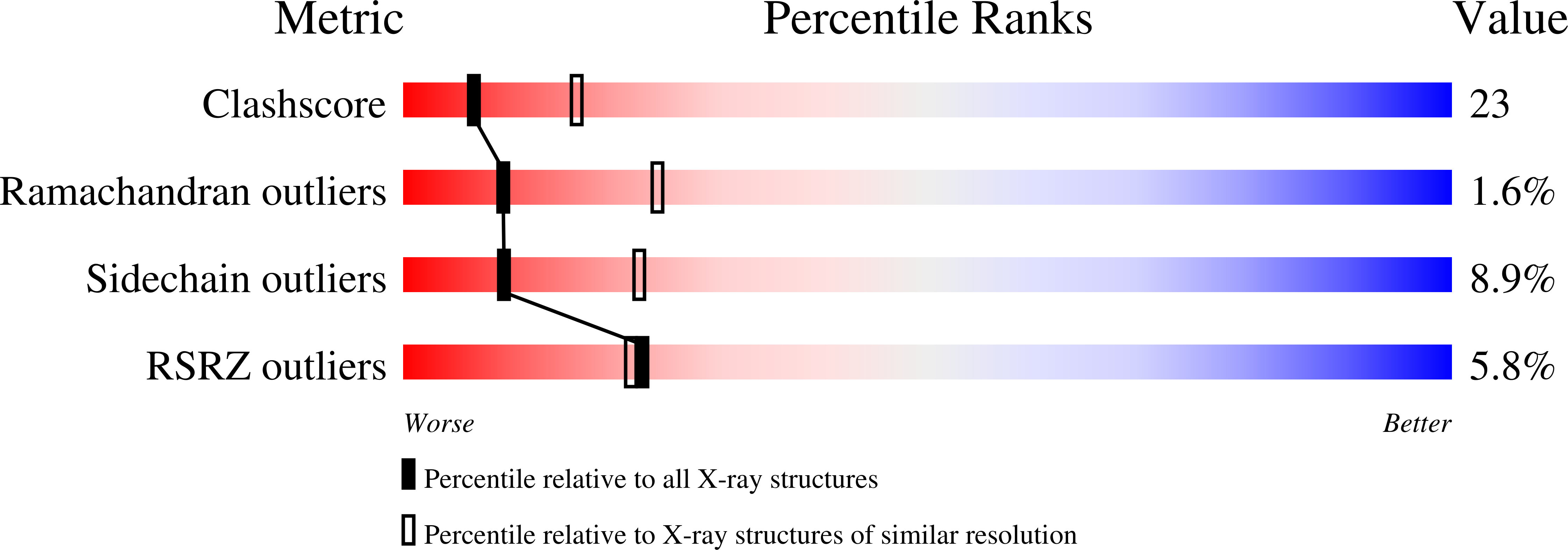
wwPDB Validation   3D Report Full Report


Ligand Structure Quality Assessment 


This is version 1.3 of the entry. See complete history


Literature

Crystal structure of pyridoxal kinase in complex with roscovitine and derivatives

Tang, L.Li, M.-H.Cao, P.Wang, F.Chang, W.-R.Bach, S.Reinhardt, J.Ferandin, Y.Galons, H.Wan, Y.Gray, N.Meijer, L.Jiang, T.Liang, D.-C.

(2005) J Biol Chem 280: 31220-31229

  • DOI: https://doi.org/10.1074/jbc.M500805200
  • Primary Citation of Related Structures:  
    1YGJ, 1YGK, 1YHJ

  • PubMed Abstract: 

    Pyridoxal kinase (PDXK) catalyzes the phosphorylation of pyridoxal, pyridoxamine, and pyridoxine in the presence of ATP and Zn2+. This constitutes an essential step in the synthesis of pyridoxal 5'-phosphate (PLP), the active form of vitamin B6, a cofactor for over 140 enzymes. (R)-Roscovitine (CYC202, Seliciclib) is a relatively selective inhibitor of cyclin-dependent kinases (CDKs), currently evaluated for the treatment of cancers, neurodegenerative disorders, renal diseases, and several viral infections. Affinity chromatography investigations have shown that (R)-roscovitine also interacts with PDXK. To understand this interaction, we determined the crystal structure of PDXK in complex with (R)-roscovitine, N6-methyl-(R)-roscovitine, and O6-(R)-roscovitine, the two latter derivatives being designed to bind to PDXK but not to CDKs. Structural analysis revealed that these three roscovitines bind similarly in the pyridoxal-binding site of PDXK rather than in the anticipated ATP-binding site. The pyridoxal pocket has thus an unexpected ability to accommodate molecules different from and larger than pyridoxal. This work provides detailed structural information on the interactions between PDXK and roscovitine and analogs. It could also aid in the design of roscovitine derivatives displaying strict selectivity for either PDXK or CDKs.


  • Organizational Affiliation

    National Laboratory of Biomacromolecules, Institute of Biophysics, Chinese Academy of Sciences, 15 Datun Road, Chaoyang District, Beijing 100101, China.


Macromolecules
Find similar proteins by:  (by identity cutoff)  |  3D Structure
Entity ID: 1
MoleculeChains Sequence LengthOrganismDetailsImage
Pyridoxal kinase312Ovis ariesMutation(s): 0 
EC: 2.7.1.35
UniProt
Find proteins for P82197 (Ovis aries)
Explore P82197 
Go to UniProtKB:  P82197
Entity Groups  
Sequence Clusters30% Identity50% Identity70% Identity90% Identity95% Identity100% Identity
UniProt GroupP82197
Sequence Annotations
Expand
  • Reference Sequence
Small Molecules
Ligands 1 Unique
IDChains Name / Formula / InChI Key2D Diagram3D Interactions
RMC
Query on RMC

Download Ideal Coordinates CCD File 
B [auth A](2R)-2-({6-[BENZYL(METHYL)AMINO]-9-ISOPROPYL-9H-PURIN-2-YL}AMINO)BUTAN-1-OL
C20 H28 N6 O
YPYWONAECUVKHY-MRXNPFEDSA-N
Experimental Data & Validation

Experimental Data

  • Method: X-RAY DIFFRACTION
  • Resolution: 2.70 Å
  • R-Value Free: 0.280 
  • R-Value Work: 0.217 
  • Space Group: P 31 2 1
Unit Cell:
Length ( Å )Angle ( ˚ )
a = 100.864α = 90
b = 100.864β = 90
c = 55.706γ = 120
Software Package:
Software NamePurpose
CrystalCleardata collection
CrystalCleardata reduction
AMoREphasing
CNSrefinement
CrystalCleardata scaling

Structure Validation

View Full Validation Report


Ligand Structure Quality Assessment 


Entry History 

Revision History  (Full details and data files)

  • Version 1.0: 2005-07-05
    Type: Initial release
  • Version 1.1: 2008-04-30
    Changes: Version format compliance
  • Version 1.2: 2011-07-13
    Changes: Version format compliance
  • Version 1.3: 2024-03-13
    Changes: Data collection, Database references, Derived calculations, Structure summary