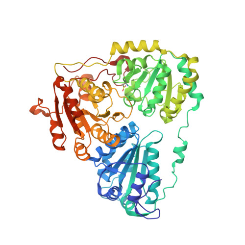
1Z8N

Crystal structure of Arabidopsis thaliana Acetohydroxyacid synthase In Complex With An Imidazolinone Herbicide, Imazaquin


Experimental Data Snapshot

  • Method: X-RAY DIFFRACTION
  • Resolution: 2.80 Å
  • R-Value Free: 0.216 
  • R-Value Work: 0.199 
  • R-Value Observed: 0.200 

Starting Model: experimental
View more details

wwPDB Validation   3D Report Full Report


Ligand Structure Quality Assessment 


This is version 1.4 of the entry. See complete history


Literature

Herbicide-binding sites revealed in the structure of plant acetohydroxyacid synthase

McCourt, J.A.Pang, S.S.King-Scott, J.Guddat, L.W.Duggleby, R.G.

(2006) Proc Natl Acad Sci U S A 103: 569-573

  • DOI: https://doi.org/10.1073/pnas.0508701103
  • Primary Citation of Related Structures:  
    1YBH, 1YHY, 1YHZ, 1YI0, 1YI1, 1Z8N

  • PubMed Abstract: 

    The sulfonylureas and imidazolinones are potent commercial herbicide families. They are among the most popular choices for farmers worldwide, because they are nontoxic to animals and highly selective. These herbicides inhibit branched-chain amino acid biosynthesis in plants by targeting acetohydroxyacid synthase (AHAS, EC 2.2.1.6). This report describes the 3D structure of Arabidopsis thaliana AHAS in complex with five sulfonylureas (to 2.5 A resolution) and with the imidazolinone, imazaquin (IQ; 2.8 A). Neither class of molecule has a structure that mimics the substrates for the enzyme, but both inhibit by blocking a channel through which access to the active site is gained. The sulfonylureas approach within 5 A of the catalytic center, which is the C2 atom of the cofactor thiamin diphosphate, whereas IQ is at least 7 A from this atom. Ten of the amino acid residues that bind the sulfonylureas also bind IQ. Six additional residues interact only with the sulfonylureas, whereas there are two residues that bind IQ but not the sulfonylureas. Thus, the two classes of inhibitor occupy partially overlapping sites but adopt different modes of binding. The increasing emergence of resistant weeds due to the appearance of mutations that interfere with the inhibition of AHAS is now a worldwide problem. The structures described here provide a rational molecular basis for understanding these mutations, thus allowing more sophisticated AHAS inhibitors to be developed. There is no previously described structure for any plant protein in complex with a commercial herbicide.


  • Organizational Affiliation

    School of Molecular and Microbial Sciences, University of Queensland, Brisbane QLD 4072, Australia.


Macromolecules
Find similar proteins by:  (by identity cutoff)  |  3D Structure
Entity ID: 1
MoleculeChains Sequence LengthOrganismDetailsImage
Acetolactate synthase590Arabidopsis thalianaMutation(s): 1 
Gene Names: ILVB
EC: 2.2.1.6
UniProt
Find proteins for P17597 (Arabidopsis thaliana)
Explore P17597 
Go to UniProtKB:  P17597
Entity Groups  
Sequence Clusters30% Identity50% Identity70% Identity90% Identity95% Identity100% Identity
UniProt GroupP17597
Sequence Annotations
Expand
  • Reference Sequence
Small Molecules
Ligands 5 Unique
IDChains Name / Formula / InChI Key2D Diagram3D Interactions
FAD
Query on FAD

Download Ideal Coordinates CCD File 
F [auth A]FLAVIN-ADENINE DINUCLEOTIDE
C27 H33 N9 O15 P2
VWWQXMAJTJZDQX-UYBVJOGSSA-N
TPP
Query on TPP

Download Ideal Coordinates CCD File 
G [auth A]THIAMINE DIPHOSPHATE
C12 H19 N4 O7 P2 S
AYEKOFBPNLCAJY-UHFFFAOYSA-O
1IQ
Query on 1IQ

Download Ideal Coordinates CCD File 
C [auth A],
D [auth A]
2-(4-ISOPROPYL-4-METHYL-5-OXO-4,5-DIHYDRO-1H-IMIDAZOL-2-YL)QUINOLINE-3-CARBOXYLIC ACID
C17 H17 N3 O3
CABMTIJINOIHOD-QGZVFWFLSA-N
NHE
Query on NHE

Download Ideal Coordinates CCD File 
E [auth A]2-[N-CYCLOHEXYLAMINO]ETHANE SULFONIC ACID
C8 H17 N O3 S
MKWKNSIESPFAQN-UHFFFAOYSA-N
MG
Query on MG

Download Ideal Coordinates CCD File 
B [auth A]MAGNESIUM ION
Mg
JLVVSXFLKOJNIY-UHFFFAOYSA-N
Modified Residues  1 Unique
IDChains TypeFormula2D DiagramParent
CSD
Query on CSD
A
L-PEPTIDE LINKINGC3 H7 N O4 SCYS
Binding Affinity Annotations 
IDSourceBinding Affinity
1IQ BindingDB:  1Z8N Ki: min: 3000, max: 4.70e+5 (nM) from 2 assay(s)
Experimental Data & Validation

Experimental Data

  • Method: X-RAY DIFFRACTION
  • Resolution: 2.80 Å
  • R-Value Free: 0.216 
  • R-Value Work: 0.199 
  • R-Value Observed: 0.200 
  • Space Group: P 64 2 2
Unit Cell:
Length ( Å )Angle ( ˚ )
a = 179.081α = 90
b = 179.081β = 90
c = 186.077γ = 120
Software Package:
Software NamePurpose
DENZOdata reduction
SCALEPACKdata scaling
CNSrefinement
CNSphasing

Structure Validation

View Full Validation Report


Ligand Structure Quality Assessment 


Entry History 

Deposition Data

Revision History  (Full details and data files)

  • Version 1.0: 2006-01-17
    Type: Initial release
  • Version 1.1: 2008-04-30
    Changes: Version format compliance
  • Version 1.2: 2011-07-13
    Changes: Derived calculations, Version format compliance
  • Version 1.3: 2023-10-25
    Changes: Data collection, Database references, Derived calculations, Refinement description
  • Version 1.4: 2024-11-06
    Changes: Structure summary